Blood, Vol. 111, Issue 7, 3343-3354, April 1, 2008
HIF-1 regulates epithelial inflammation by cell autonomous NF
 B activation and paracrine stromal remodeling
Blood Scortegagna et al. 111: 3343
Supplemental materials for: Scortegagna et al
Files in this Data Supplement:
- Table S1. Sequences of PCR primers and TaqMan probes (JPG, 85.9 KB) -

- Figure S1. Comparison between K14-HIF-1αΔODD and K14-HIF-1α DPM mice (JPG, 75.4 KB) -
Human HIF-1 mRNA (Panel A) and total HIF-1 protein (Panel B) are significantly increased in K14-HIF-1
 ODD transgenic keratinocytes (ODD) compared to the point mutant K14-HIF-1DPM transgenic keratinocytes (DPM). Real-time RT–PCR analysis reveals a significant induction of the HIF-1 target genes, VEGF-A (Panel C) and PlGF (Panel D) in K14-HIF-1DPM compared to K14-HIF-1ODD cells. Quantification of CD45 cells did reveal a statistically significant 0.25-fold elevation in K14-HIF-1ODD transgenic mice compared to nontransgenic controls (NTG), markedly less compared to the enhanced baseline accumulation in the point mutant counterparts (Panel E). Intraepidermal neutrophilic abscesses were present six hours post-TPA challenge in K14-HIF-1ODD ears, however, they were smaller compared to intraepidermal microabscesses in TPA challenged K14-DPM transgenic mice; H&E stain (Panel F). Error bars are mean ± SEM. Results are representative of three independent experiments. Bar = 100 µm.
- Figure S2. Inhibition of PKC with the pan-specific PKC inhibitor GF09203X (GF) failed to alter the elevated level of TNFα in transgenic cells (JPG, 77.8 KB) -
Real-time RT–PCR analysis of TNF expression from total RNA extracted from transgenic (DPM) primary keratinocytes. Error bars are mean ± SEM. Results are representative of three independent experiments.
- Figure S3. PlGF immunodepletion was unable to prevent the Munro abscess formation (JPG, 85.3 KB) -
A single dose of TPA (2.5 µg) was applied on each side of the ears of transgenic mice pretreated with PlGF neutralizing antibodies. Transgenic ears were collected 6 hours later and stained with H&E. Bar = 100 µm.
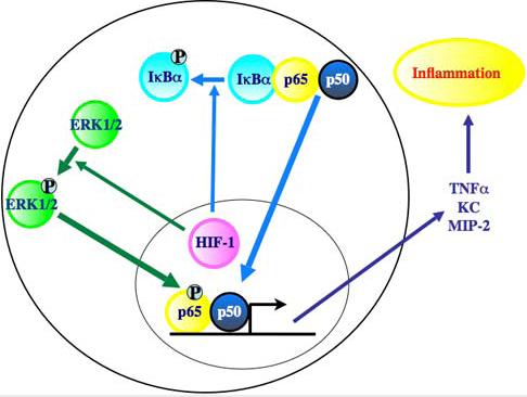
- Figure S4. Summary cartoon of HIF-1 mediated signaling to the NFκB pathway (JPG, 28.1 KB) -
HIF-1 induced NF B activation is composed of two discrete elements, phosphorylation of Ser276 on p65 and IB phosphorylation. The cartoon shows increased ERK1/2 phosphorylation by HIF-1 upregulation and consequent increase in Ser276 phosphorylation on p65. Moreover HIF-1 gain of function leads to increased IB phosphorylation through a yet unknown mechanism. Amplification of NFB transcriptional activation is responsible for the inflammatory hyperresponsiveness observed in K14-HIF-1DPM mice.