[View Larger Version of this Image]
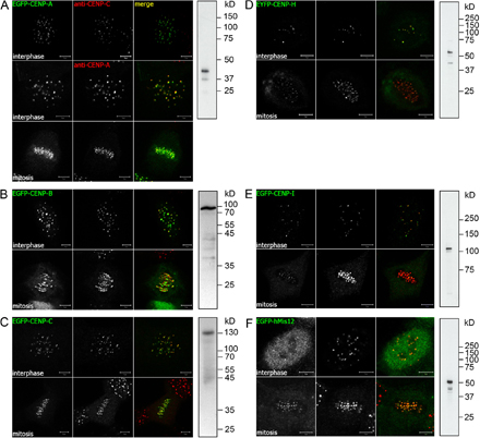
Figure S1. Characterization of GFP-tagged centromere proteins in HEp-2 cells. HEp-2 cells expressing GFP–CENP-A (A), GFP–CENP-B (B), GFP–CENP-C (C), YFP–CENP-H (D), GFP–CENP-I (E), or GFP-hMis12 (F) were analyzed by indirect immunofluorescence and confocal microscopy in interphase and mitosis as indicated. One midnucleus confocal section displaying GFP signals (left), anti–CENP-C or anti–CENP-A staining (middle), and the merged image (right) are shown. Anti-GFP Western blot strips of whole cell lysates are shown on the right of the confocal images. Bars, 5 µm.