[View Larger Version of this Image]
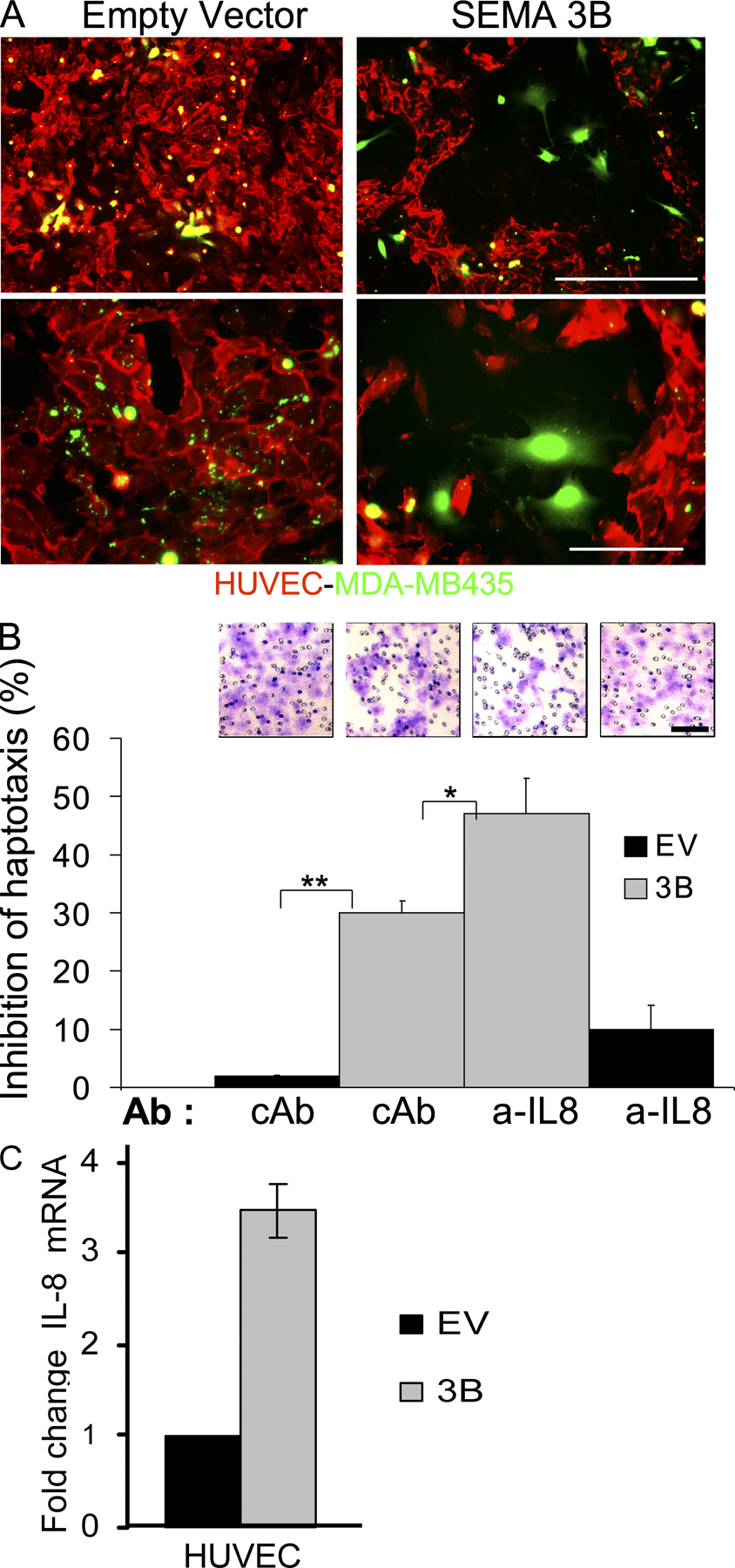
Figure S6. SEMA3B-repelling activity on endothelial cells. (A) Equal numbers of MDA-MB435 tumor cells transduced to express SEMA3B or EV control were seeded on a monolayer of HUVECs grown on glass cover slips, and co-cultured for 24 h. Before seeding, tumor cells had been labeled with Vybrant CellTracker green, whereas HUVECs were revealed by immunostaining with α-CD31 (red) at the end of the experiment. Top images were taken at low magnification, whereas bottom images were taken at high magnification. Bars: (top) 60 µm; (bottom) 20 µm. (B) Inhibition of the haptotactic migration of HUVECs through Transwell inserts in response to conditioned medium containing SEMA3B or EV control, added in the lower chamber, and in the presence of IL-8 blocking antibodies (MAB208) or control antibody (anti-CD19). The graph shows the percent inhibition of spontaneous migration, calculated by counting migrated cells stained with crystal violet (values are the mean ± SD). It has been previously shown that endothelial cell survival is sustained by autocrine secretion of IL-8 (reference 4). Notably, in the presence of IL-8–neutralizing antibodies, HUVEC migration was further inhibited. (top) Images show representative fields for each experimental condition. Bar, 60 µm. (C) The expression levels of IL-8 were measured by real time RT-PCR in the RNA of HUVECs transduced to express SEMA3B. The housekeeping gene β-actin provided the internal reference. Values indicate the fold induction of IL-8 levels in SEMA3B-treated cells versus controls, and data are the mean ± SD. *, P < 0.05.