[View Larger Version of this Image]
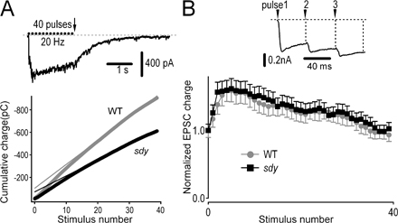
Figure S3. Smaller RRP size in sdy hippocampal CA1 pyramidal neurons. (A) Estimation of RRP size using high-frequency stimulation. (top) Representative EPSCs (20 Hz, 40 pulses) recorded from a hippocampal neuron. The arrow indicates the end of the stimulus train. (bottom) Cumulative EPSC charge values during 40 stimuli. Data points through the steady-state portion of EPSCs were fitted by linear regression and back-extrapolated to time 0 to estimate the RRP size. RRP sizes were 105.3 ± 23.6 pC in WT (gray) and 72.8 ± 14.5 pC in sdy (black; n = 13 WT neurons and 12 sdy neurons; P < 0.001). (B) The kinetics of replenishment of RRP in sdy cells were similar to those in WT cells. The normalized EPSC charge per pulse was calculated from WT (gray) or sdy (black) neurons. (inset) EPSC of three consecutive stimulation pulses. The synaptic responses were evoked by a 0.20-ms current injection (500 µA). 10 µM bicuculline was added to block GABA receptor-mediated currents. Error bars indicate the mean ± SEM.