[View Larger Version of this Image]
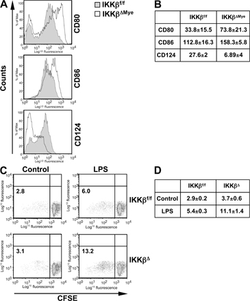
Figure S1. Increased immunostimulatory activity of IKKβΔ macrophages. (A) IKKβΔMye and IKKβf/f mice were challenged i.p. with 5 × 10⁷ CFUs GBS in PBS, and peritoneal macrophages were collected after 4 d for FACS analysis of CD86, CD80, and CD124 expression on CD11b+ macrophages. Representative histograms are shown. (B) Tabulated data of mean fluorescence intensity (MFI) of CD86, CD80, or CD124 on CD11b+ macrophages from GBS-challenged mice (data are represented as mean ± SEM of n = 6). (C) Allogenic T cell proliferation measured as CFSE dilution by FACS. CD4+ lymph node cells were purified from BALB/c mice and labeled with CFSE. 5 × 10⁵ T cells were co-cultured with 6 × 10³ IKKβf/f or IKKβΔ macrophages on the C57Bl6/J background in triplicate with and without LPS stimulation. CFSE dilution was measured after 4 d, and 7-AAD staining was used to exclude nonviable cells from analysis. Representative dot plots are shown indicating the percentage of proliferating cells in the bottom left quadrant. (D) Tabulated data of mean ± SEM of n = 3 independent experiments.