Supplementary material for Calhoun et al. (1999) Proc. Natl. Acad. Sci. USA 96 (24), 14088-14093.

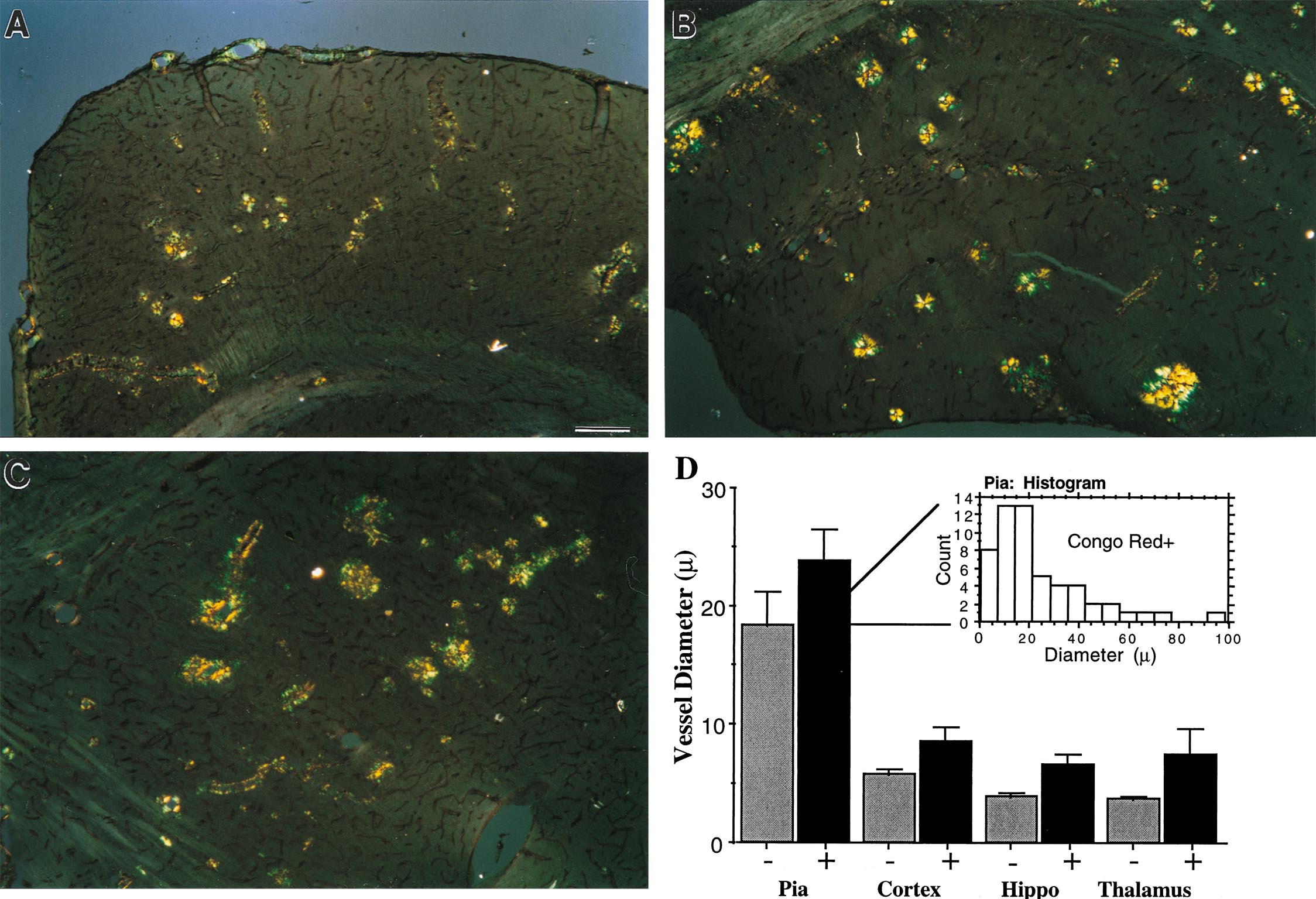
Fig. 7.
Quantification of amyloid-positive vessel diameter and surface area on ß-dystroglycan and Congo red double-stained sections. (A-C) ß-dystroglycan immunolabeling was used to visualize all vessels (brown reaction product), and Congo red was used to visualize CAA (birefringence) in neocortex (A), hippocampus (B), and thalamus (C). Percent of Congo red-positive vessel surface area to total ß-dystroglycan labeled surface area was quantified by using stereological techniques, and results are shown in Table 1 in the main text. (D) The mean inner diameter of vessels with vascular amyloid (+) and vessels free of amyloid (–) was measured. Affected vessels had a larger diameter than vessels free of amyloid in each of the four regions analyzed. The size histogram of affected pial vessels (Inset) indicates, however, that fibrillary amyloid was present in vessels of all sizes. The size distribution of affected vessels in the other regions analyzed also included the full range of vessel diameters. Widespread amyloid deposition such as that shown in this figure occurred in homozygous animals at approximately 16 months of age. (Bar in A is 150 µm for A-C.)