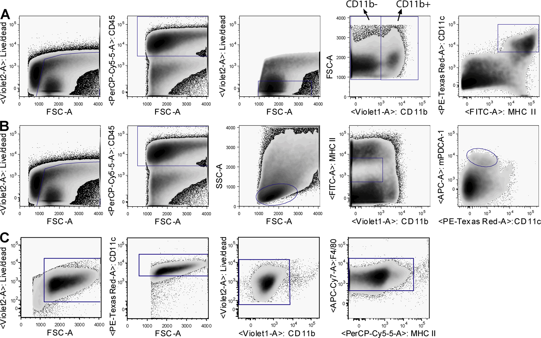
[View Larger Version of this Image]
Figure S2. Gating strategies in an eight-color flow cytometric analysis of lung samples. (A) Myeloid DCs were gated as live cells (CD45+ and low fluorescent). Next, CD11b+ and CD11b− fractions were determined, and within these populations the MHCII+CD11c+ DCs were gated. (B) pDCs were gated as live cells (CD45+ in the lymphocyte gate). They were then gated as CD11b−MHCIIint cells expressing CD11cint and high mPDCA-1. (C) Alveolar macrophages were determined in BAL fluid as highly autofluorescent cells expressing high levels of CD11c. The CD11b− fraction was then gated for F4/80+ cells.