[View Larger Version of this Image]
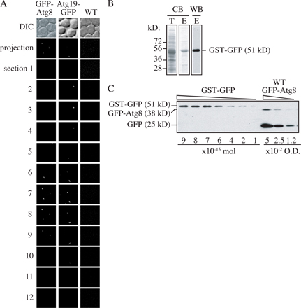
Figure S1. Examination of the relationship between GFP-tagged protein amount and fluorescence intensity. (A) Wild-type (SEY6210) and Atg19-GFP (JGY018) cells cultured in nutrient-rich medium and GFP-Atg8 (YZX247) cells starved for 2 h were subjected to microscopy. A stack of 12 Z-section pictures was collected and projected into a 2D image, and the fluorescence signals per cell were quantified as described in Materials and methods. The intensity values from wild-type cells were subtracted as background. Bar, 2 µm. (B) The GST-GFP fusion protein was purified from E. coli BL21 cells as a protein standard as described in Materials and methods. Total cell lysate and elution of glutathione Sepharose were examined by Coomassie Blue staining. The elution was confirmed by Western blot using anti-YFP antibody. (C) Quantitative Western blot. Different amounts of purified GST-GFP were loaded as standard, and protein samples from other GFP-tagged strains (GFP-Atg8 is shown here) were tested on the same gel. Duplicate samples of GFP-Atg8 corresponding to 0.05, 0.025, and 0.0125 OD600 units of cells were loaded to fit both GFP-Atg8 and free GFP bands into the range of the standard protein.