[View Larger Version of this Image]
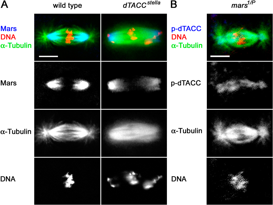
Figure S3. Distribution of Mars and p-dTACC in embryos laid by dTACC and mars mutant mothers, respectively. (A) Mars localization is not disrupted in a dTACC mutant background. Wild-type or dTACCstella mutant embryos were fixed and stained for Mars (blue) DNA (red) and α-tubulin (green). Mars staining was found on the poles of spindle MTs in both wild-type and dTACC mutant embryos. (B) Example of a normal-looking spindle from a mars1/P embryo, visualized by DNA (red) and α-tubulin staining (green). p-dTACC staining (blue) is visible on both the centrosome and the spindle.