Blood, Vol. 112, Issue 3, 626-634, August 1, 2008
TGF-β signaling in thymic epithelial cells regulates thymic involution and postirradiation reconstitution
Blood Hauri-Hohl et al. 112: 626
Supplemental materials for: Hauri-Hohl et al
Files in this Data Supplement:
- Table S1. Accumulation of mature SP thymocytes in TGFβRIIlox/lox::Foxn1-Cre mice (PDF, 41.4 KB) -
Measurement of total and mature CD4 and CD8 T cells (the latter being TCRhigh CD24low). Results are given as the percentage of total thymocytes (% of total) and as absolute cell numbers (×106) comparing TGF RIIlox/lox::Foxn1-Cre and TGFRIIlox/lox littermates of different ages. Representative experiments with ≥ 3 animals per group are shown; p-values obtained using a two-sided t-test.
 - Figure S1. Quantification of early thymocyte precursors in TGFβRIIlox/lox::Foxn1-Cre and TGFβRIIlox/lox mice (JPG, 33 KB) -
(A) Frequency of ETP (lin−CD25−CD44+c-kit+Sca-1+) in thymi of young (8weeks) and old (64 weeks) mice. (B, C) Cellularity of the different DN subpopulations in thymi of 8-week-old TGFRIIlox/lox::Foxn1-Cre and TGFRIIlox/lox mice.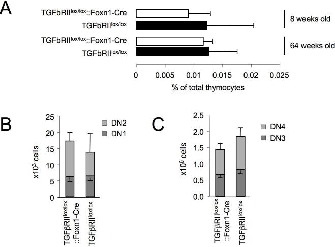
- Figure S2. TGF-β and TGFβRII expression in the ageing thymus (JPG, 30.8 KB) -
(A) TGF-1 and TGF-2 mRNA and (B) TGF- protein expression in thymi of naïve mice at 2, 5 and 24 months of age. TGF1 and TGF2 expression was quantified by qRT-PCR using the following primer pairs: TGF1 forward: 5′-GACCGCAACAACGCCATCTA-3′, TGF1 reverse: 5′-GGCGTATCAGTGGGGGTCAG-3′; TGF2 forward: 5′-TTTCAGCCTTTTCTGCGTCA-3′; TGF2 reverse: 5′-CTACATTTGTGCGAACTTCTGTGTT-3′. Amplicons were normalized to GAPDH (forward 5′-GGTGAAGGTCGGTGTGAACG-3′, reverse 5′-ACCATGTAGTTGAGGTCAATGAAGG-3′). For protein detection, equal amounts of total thymic protein were separated by SDS-PAGE under non-reducing conditions and blotted on a nitrocellulose membrane as described previously (Torrealba et al., J Immunol. 2004; 172:5753-5764). TGF- was detected using a pan-TGF antibody (Clone 1D11, R&D Systems Inc.). (C) Expression of TGFRII in sorted TEC derived from 5 month old naïve mice (TGFRII forward 5′-CAGTGTGCTGAGAGACCGAG-3′, TGFRII reverse 5′-AGCACTCGGTCAAAGTCTCA-3′).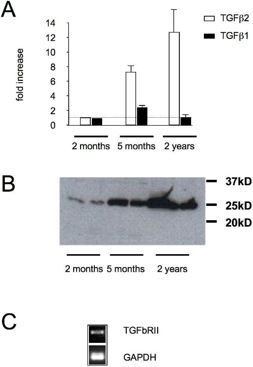
- Figure S3. Thymic export and TCR diversity among peripheral T cells of aged TGFβRIIlox/lox::Foxn1-Cre and TGFβRIIlox/lox mice (JPG, 55.2 KB) -
(A) Determination of CD44low (i.e. naïve) splenic T cells in 28 week old TGFRIIlox/lox::Foxn1-Cre and TGFRIIlox/lox mice. (B) TCR V diversity among splenic T cells of 1 year old TGFRIIlox/lox::Foxn1-Cre and TGFRIIlox/lox mice For this purpose, the mouse V TCR Screening Panel kit (Cat #557004, BD Bioscience, Allschwil, Switzerland) was used. One representative experiments (≥4 animals per group) out of two is shown; p-value was obtained using a t-test. (C) Measurement of sjTREC in splenic T cells from aged (52 weeks old) TGFRIIlox/lox::Foxn1-Cre and control mice. The data is representative of two independent experiments using methods, as previously described (Krenger et al., J Immunol. 2004, 172: 7359–7367).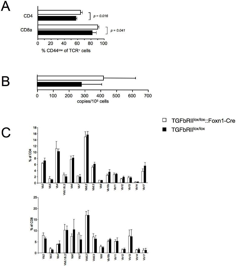
- Figure S4. The proliferative response of TECs to TGF-β (JPG, 36 KB) -
Cortical (TEC1.2, TEC1.4) and medullary (TEC2.3, TECC6) TEC lines were grown in the presence (10ng/ml) or absence of hTGF-1 for 48 hours. (A) Cell recovery after stimulation period. The dotted line denotes the input cell number. (B) 3H-thymidine incorporation during the last 16 hours of culture.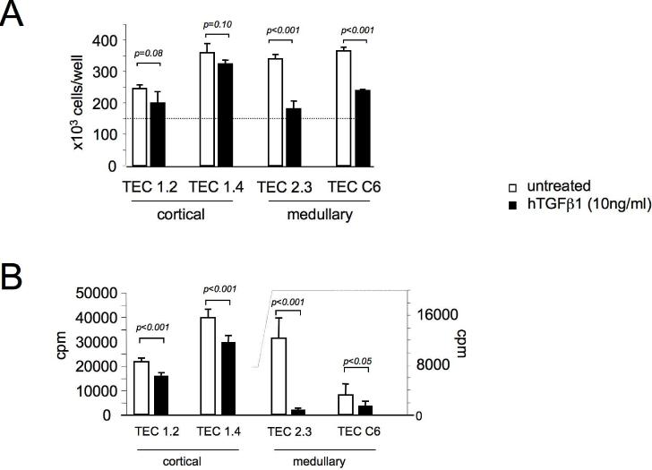
- Figure S5. Thymic morphology of aged TGFβRIIlox/lox::Foxn1-Cre and TGFβRIIlox/lox mice (JPG, 120 KB) -
(i–iv) Representative thymus tissue sections from 52 week old mutant and control mice using H+E staining. Comparable cortico-medullary demarcation was noted (Magnification: 4× in i and ii, 10× in iii and iv). (v–viii) Immunohistochemistry of thymic tissue isolated from 52 week old wildtype and mutant mice for the expression of epithelial markers (K5: medulla; K18: cortex) and fibrobasts (ERTR7). Note an equivalent stromal composition in the medulla and at the cortico-medullary junction (Magnification: 20× in v–viii).
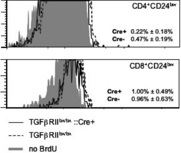
- Figure S6. Proliferation of mature thymic T cells (JPG, 31.5 KB) -
In vivo proliferation of mature (TCRhigh, CD24low) thymic T cells, measured by incorporation of BrdU 1.5 hours after i.p. injection of 1mg of BrdU.