[View Larger Version of this Image]
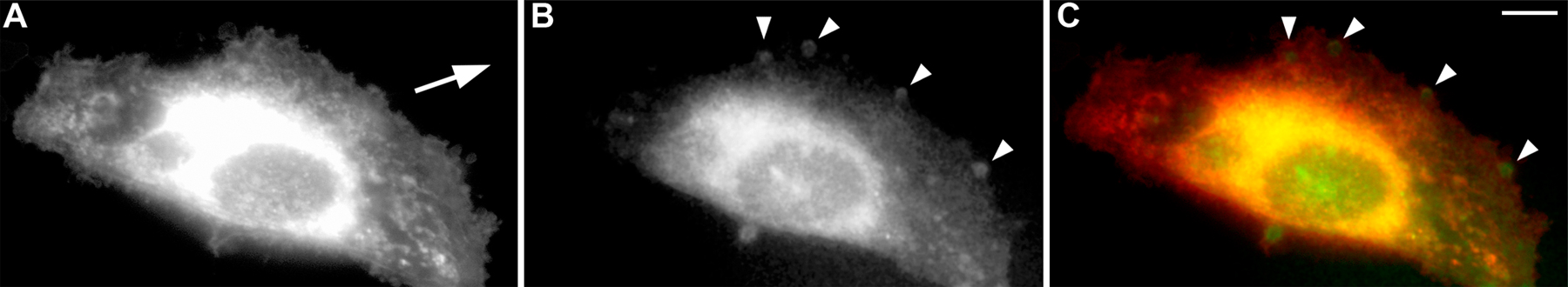
Figure S1. Validation that Gαi3 is preferentially localized within pseudopods at the leading edge during cell migration using DiD as a PM marker. HeLa cells were transfected with Gαi3-YFP and grown to confluence on coverslips for 36 h before induction of migration by scratch wounding. Cells were then treated with 5 mg/ml DiD in ethanol to a final concentration of 3.3 µg/ml and incubated for 8 min at 37°C before fixation. Representative images of a single migrating cell (arrow shows direction of migration) are displayed as analyzed through the red (A; DiD-stained membranes) and green (B; Gαi3-YFP) channels, whereas C displays the merged image. As expected, DiD labels intracellular membranes (perinuclear region and vesicular structures). However, DiD (red)/YFP (green) ratios for fluorescent signals at the PM show that Gαi3 accumulates (blushes previously seen in Fig. 2 A) within pseudopods (arrowheads) at the leading edge, whereas DiD is more uniformly distributed. Bar, 10 µm.