[View Larger Version of this Image]
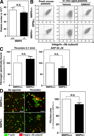
Figure S2. Gene deletion of MMP-9 did not prevent GPIbα shedding in aged platelets and did not change platelet function mediated through integrin αIIbβ3. (A) Peripheral blood from MMP9−/− mice or their control MMP9+/+ mice was obtained from a retroorbital venous plexus, and platelet numbers were counted using an automated counter (n = 7 each). (B) Washed platelets obtained from MMP9+/+ mice or MMP9−/− mice were incubated at 37°C for 1 d in the presence of GM6001 or a control, 1% DMSO. Representative flow cytometry dot plots for freshly washed platelets or in vitro–injured platelets illustrate the expression of GPIbα and the αIIb integrin subunit. (C) The graph shows the specific fibrinogen binding to αIIbβ3 stimulated with 0.1 U/ml thrombin or 50 µM adenosine diphosphate. The value of bound in MMP9+/+ platelets is defined as 100%. (D) Representative fluorescence images of platelet spreading in MMP9+/+ or MMP9+/+ platelets. All samples were stained with Alexa 488 phalloidin to mark F-actin (green) and with anti-αIIb integrin subunit antibody followed by Alexa 567 (red). Bar, 10 µm. The graph summarizes the surface area of platelet spreading quantified by National Institutes of Health image software. Results are the mean ± SEM from three independent experiments.