[View Larger Version of this Image]
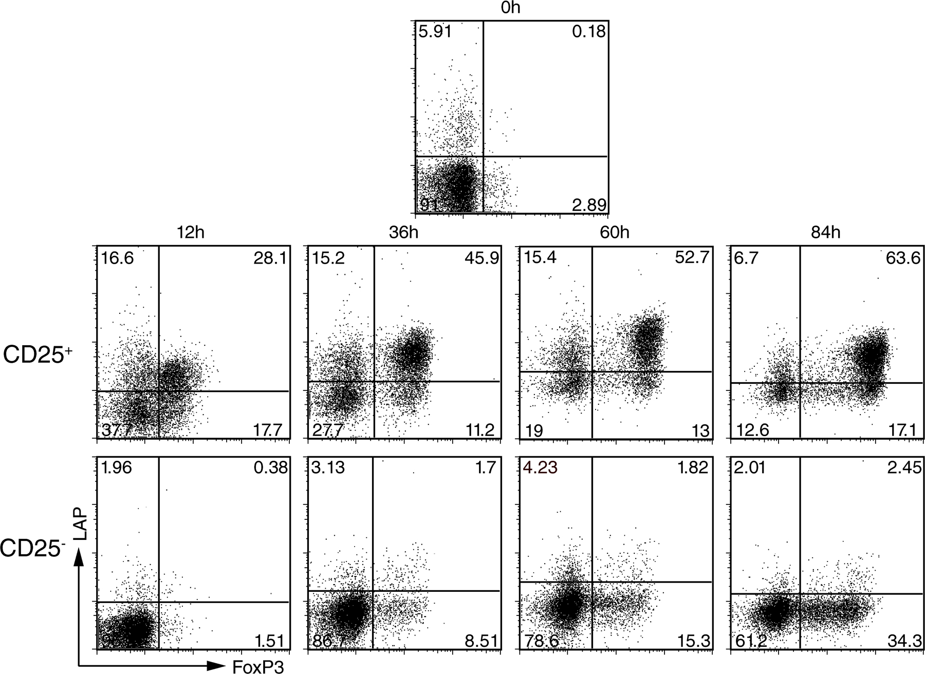
Figure S1. (A) CD4+ T cells from human peripheral blood were analyzed for expression of LAP and FoxP3. (B) AutoMACS-purified CD4+CD25+ (∼50% FoxP3+) and CD4+CD25− T cells from human peripheral blood were activated with anti-CD3 and IL-2 and analyzed for FoxP3 and LAP expression at the indicated time points. One representative experiment out of three is shown.