[View Larger Version of this Image]
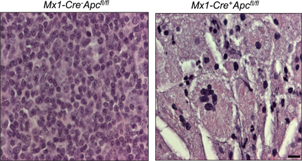
Figure S4. Histological analysis of thymus from Mx1-Cre−Apcfl/fl primary mice (left) and Mx1-Cre+Apcfl/fl primary mice (right) 4 d after three doses of pI-pC. Sections were stained with hematoxylin and eosin. Bar, 10 µm.