[View Larger Version of this Image]
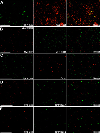
Figure S1. Fluorescence analyses in DMZ cells of Xenopus laevis embryos. Embryos were microinjected into the two dorsal blastomeres at the four-cell stage with a combination of the indicated mRNAs (500 pg GFP Dsh, 500 pg myc Dsh, 500 pg myc Fz7, 500 pg myc Rab5, 500 pg GFP Rab5, 10 ng xßarr2 MO, 500 pg GFP hCav-1, and 500 pg GFP mCav-2). DMZ explants were dissected at stage 11–11.5 and then subjected to fluorescence analysis. (A) Dsh colocalized with Rab5. (B) xßarr2 MO inhibited colocalization between Fz7 and Rab5. (C–E) Dsh did not colocalize with Cav-1 (C and D) and Cav-2 (E) in CE movements. Immunostaining in C used anti–hCav-1 antibodies. Bars, 20 µm.