[View Larger Version of this Image]
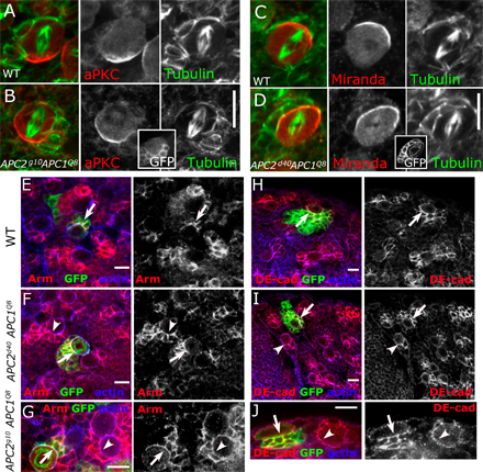
Figure S3. APC mutant NBs exhibit normal polarity third instar larval brains. Antigens are indicated. (A and B) NBs fixed and stained for the apical marker aPKC and tubulin. (A) Wild-type NB. (B) APC2g10 APC1Q8 clone. (C and D) NB fixed and stained for the basal marker Miranda and tubulin. (C) Wild-type NB. (D) APC2d40 APC1Q8 clone. The insets are images of GFP::CD8, which positively marks the NB clones. (E–J) Clonal NBs are indicated by GFP staining (arrows), whereas adjacent wild-type NBs are indicated by arrowheads. (E–G) NBs fixed and stained for Armadillo (Arm) and actin. (H–J) NBs fixed and stained for Drosophila E-cadherin (DE-cad) and actin. (E and H) Control clones. (F and I) APC2d40 APC1Q8 clones. (G and J) APC2g10 APC1Q8 clones. Bars, 10 µm.