[View Larger Version of this Image]
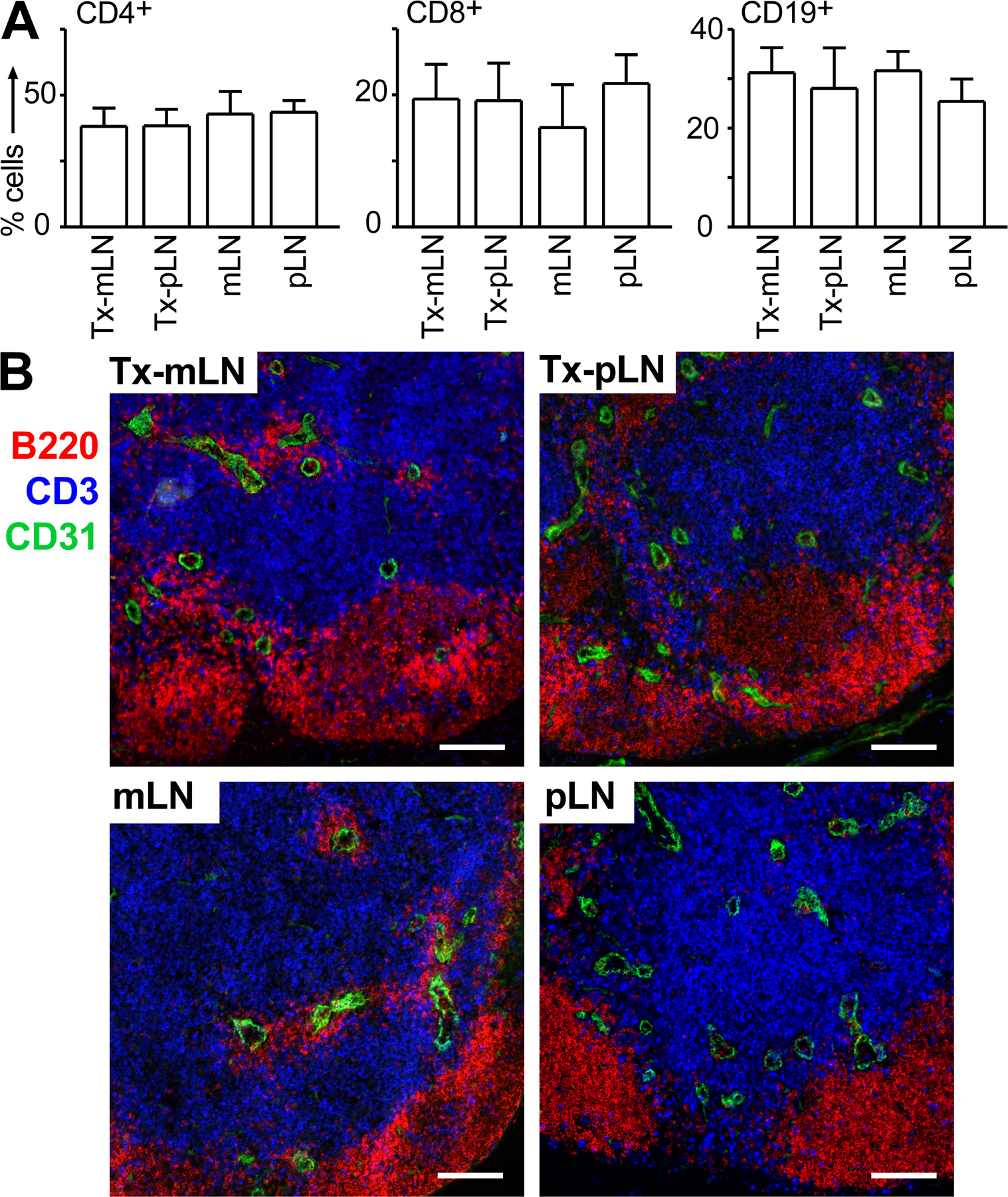
Figure S1. Transplanted LN display normal cellular composition and LN architecture. The endogeneous mLN was excised and replaced by either pLN or mLN fragments isolated from WT mice. 8–10 wk after surgery, Tx-pLN and Tx-mLN were isolated and their cellular composition and LN architecture analyzed. (A) Frequencies of CD4+ and CD8+ T cells, as well as CD19+ B cells, were comparable between Tx-mLN, Tx-pLN, and mLN and pLN isolated from nonmanipulated WT mice. Data depict the mean and SD of four mice per group analyzed in two independent experiments. (B) Cryosections of Tx-LN and endogenous LN were stained with anti-B220 (red), anti-CD3 (blue), and anti-CD31 (green). Tx-LN displayed normal segregation of T and B cell zones and unaltered numbers and distribution of CD31-expressing vessels. Tx-mLN, as well as Tx-pLN, showed slightly increased frequencies of germinal centers compared with LN isolated from nonmanipulated mice (not depicted). Bars, 100 µm.