Blood, Vol. 112, Issue 12, 4755-4764, December 1, 2008
Rapidly proliferating CD44hi peripheral T cells undergo apoptosis and delay posttransplantation T-cell reconstitution after allogeneic bone marrow transplantation
Blood Alpdogan et al. 112: 4755
Supplemental materials for: Alpdogan et al
Files in this Data Supplement:
- Figure S1. Peripheral donor CD4 and CD8 T cells recover slowly after allogenic T-cell–depleted bone marrow transplant (JPG, 46.6 KB) -
Lethally irradiated (1,100 cGy) 8–10-week-old LP mice were transplanted with 5 × 10⁶ T-cell–depleted B6 BM, and numbers of donor splenic CD4 and CD8 T cells (Ly9.1¬negative due to the allelic difference between the B6 and LP strains) over time is shown. Horizontal red line indicates the number of cells in normal LP mice.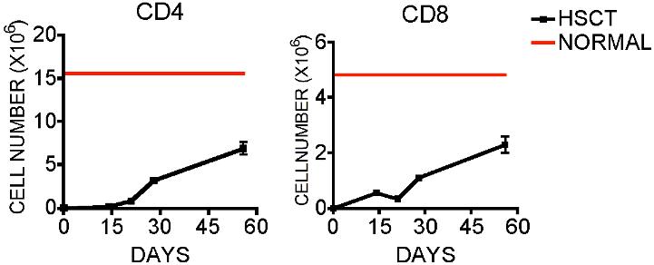
- Figure S2. Infused donor T cells rapidly expand in irradiated host animals (JPG, 119 KB) -
Lethally irradiated (1,100cGy) LP mice were transplanted with 5 × 10⁶ T-cell–depleted B6 BM with or without 0.5 × 10⁶ B6 Ly5.1 T cells and harvested at days 7, 14 and 21. (A) Total splenic cellularity and cellular ontogeny of mice transplanted with BM and 0.5 × 10⁶ B6 Ly5.1 T cells was determined as described in the results. (B–C) Weight change and mortality of animals post-transplant is shown. (N=10 in group receiving T cells).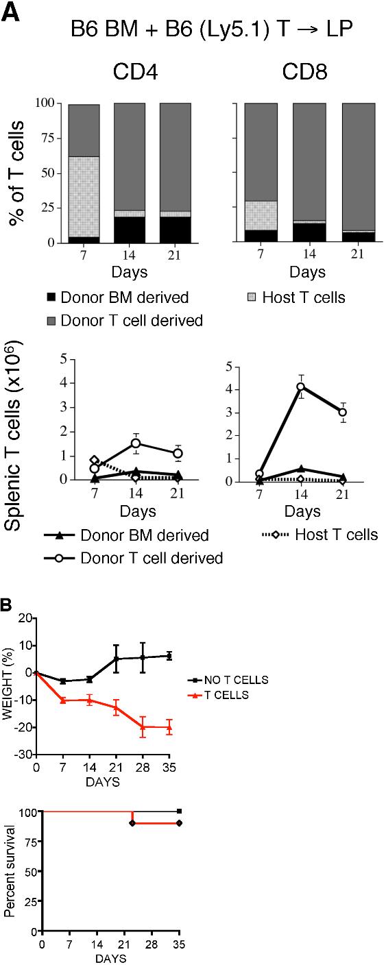
- Figure S3. GVHD does not affect the inability of Bcl-2 overexpression or Fas deficiency to modulate peripheral donor-derived T-cell apoptosis after T-cell replete BMT (JPG, 80.6 KB) -
Lethally irradiated (1,100 cGy) LP mice were transplanted with either 5 × 10⁶ B6 (WT) or lpr BM and 0.5 × 10⁶ B6 Ly5.1 T cells. (A) Total thymic cellularity was determined (B–C) apoptosis of CD4+ and CD8+ donor T cells in the BM and spleen are shown. Lethally irradiated (1,100 cGy) LP mice were transplanted with either 5 × 10⁶ B6 (WT) or Bcl-2 Tg BM and 0.5 × 10⁶ B6 Ly5.1 T cells. (D) Total thymic cellularity was determined. * denotes p+ and CD8+ donor T cells in the BM and spleen are shown.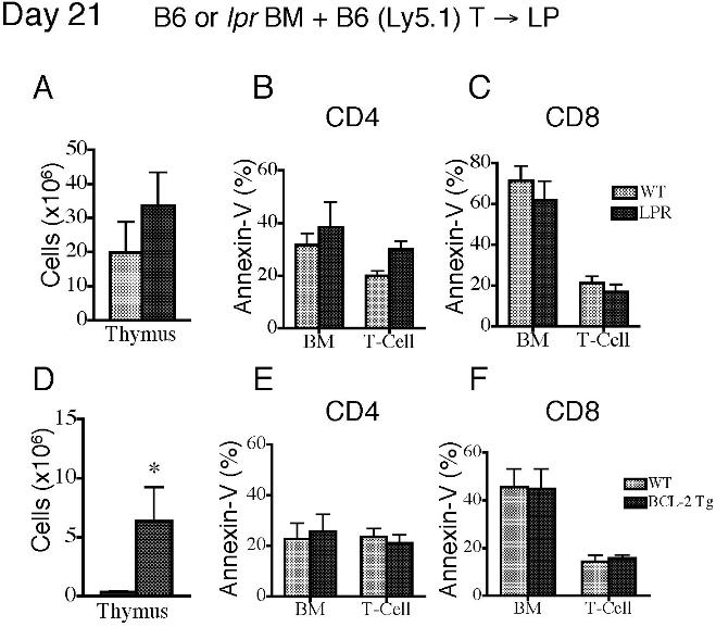
- Figure S4. DP, SP-CD4 and SP-CD8 cells in the thymus of Rag-2-eGFP Tg mice are eGFPhi (JPG, 107 KB) -
CD44hi CD4+ and CD8+ T cells are more apoptotic than CD44lo T cells regardless of recent thymic emigrant status. (A) Lethally irradiated (850 cGy) BALB/c recipients received 5 × 10⁶ T-cell–depleted Rag-2-eGFP Tg BM and were sacrificed on day 42. Thymocytes were analyzed by flow cytometry. (B–C) Splenocytes from non-transplanted 8-week-old and 8-month-old Rag-2-eGFP Tg mice were analyzed. Both eGFPloCD44hi and eGFPhiCD44hi CD4+ and CD8+ T cells display an elevated level of apoptosis as measured by annexin-V staining.
- Figure S5. Alloreactive fast-proliferating T cells have a high level of apoptosis (JPG, 73.4 KB) -
(A–B) Lethally irradiated (1,100 cGy) B6 Ly5.1 and BALB/c (850 cGy) recipients were transplanted with 2 × 10⁷ CFSE-labeled B6 purified T cells and harvested after 3 days and analyzed for apoptosis by annexin-V staining. Donor T cells are shown.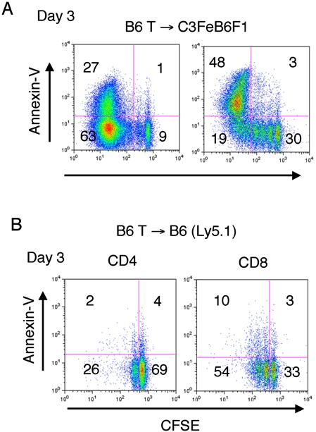
- Figure S6. Rapid spontaneously proliferating non-alloreactive adoptively-transferred donor T cells acquire a partially activated phenotype (JPG, 93.8 KB) -
Lethally irradiated (1,100 cGy) B6 Ly5.1 recipients were transplanted with 2 × 10⁷ CFSE-labeled B6 purified T cells and harvested after 7 days. Donor T cells and their expression of CD25, CD44, and CD62L are shown.
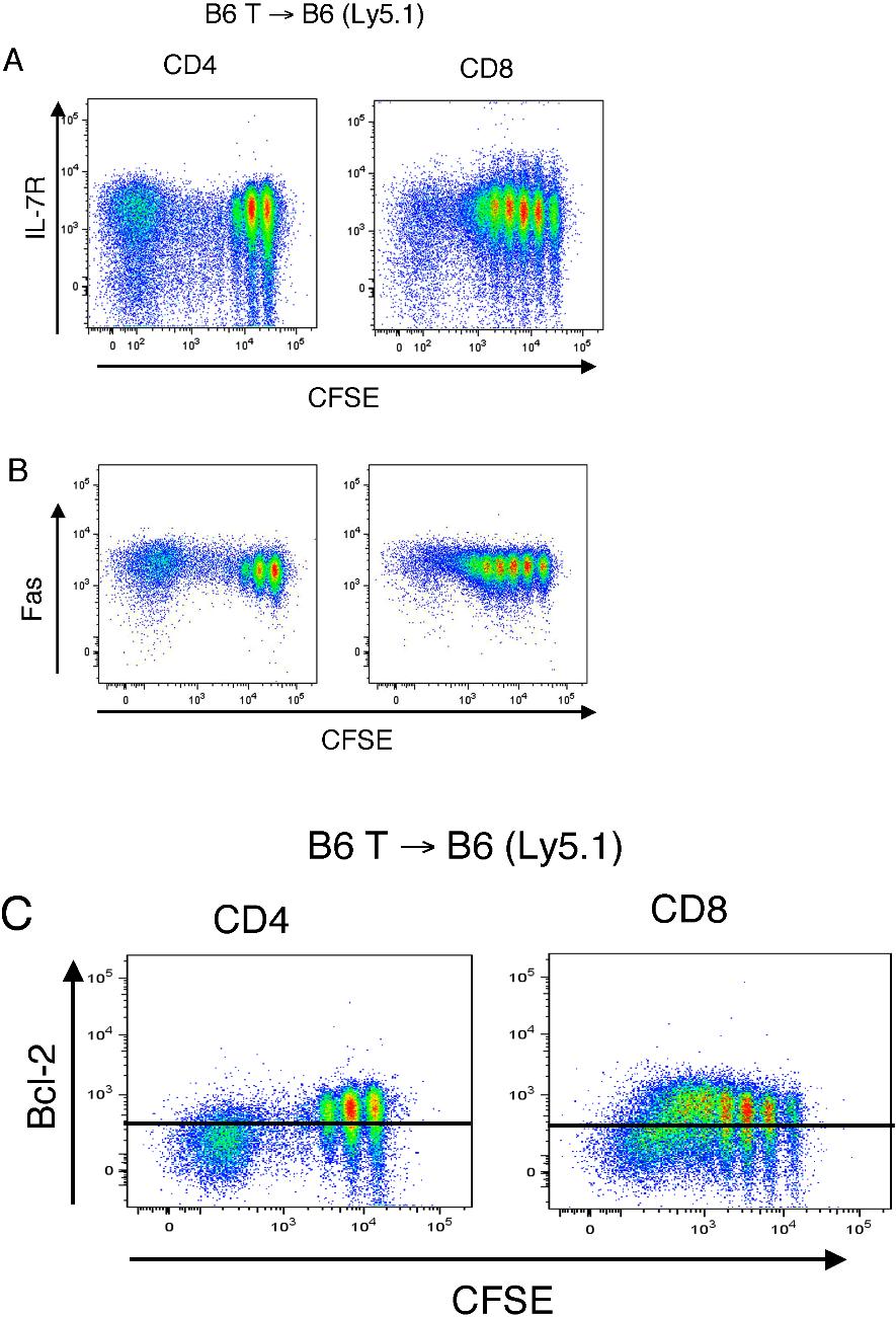
- Figure S7. Rapid spontaneously proliferating non-alloreactive adoptively-transferred donor T cells have decreased expression of Bcl-2 but not IL-7R or Fas (JPG, 184 KB) -
Lethally irradiated (1,100 cGy) B6 Ly5.1 recipients were transplanted with 2 × 10⁷ CFSE-labeled B6 purified T cells. (A) IL-7Rα expression on CD4+ and CD8+ T cells was determined at day 7. Plots show representative data from three independent experiments. (B) Fas expression on CD4+ and CD8+ T cells was determined at day 7. Data from two independent experiments. (C) Intracellular Bcl-2 expression was determined at day 7. Horizontal lines represent the limits of isotype control. Plots show representative data from two independent experiments.