Blood, Vol. 112, Issue 13, 5007-5015, December 15, 2008
Genetic perturbation of the putative cytoplasmic membrane-proximal salt bridge aberrantly activates  4 integrins
Blood Imai et al. 112: 5007
Supplemental materials for: Imai et al
Files in this Data Supplement:
- Document 1. Supplemental materials and methods (PDF, 67.1 KB)
- Table S1. α4 integrin expression on donor splenic lymphocytes (PDF, 211 KB)
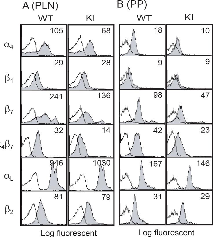
- Figure S1. Cell-surface expression of integrins on lymphocytes from peripheral lymph nodes (A) and Peyer’s patches (B) (JPG, 96.3 KB) -
Numbers denote mean fluorescent intensity (MFI). Background binding of isotype control antibodies is shown with open histograms.
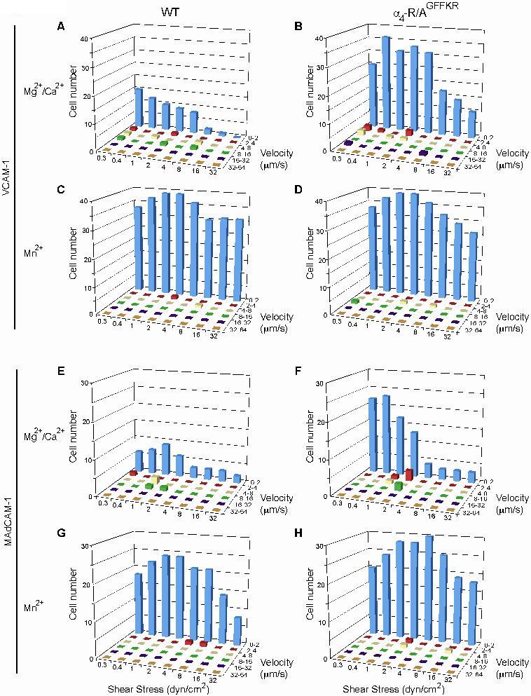
- Figure S2. Enhanced adhesive interactions of α4−R/AGFFKR splenocytes with VCAM-1 and MAdCAM-1 under shear stress (JPG, 174 KB) -
Wild-type (WT; A, C, E, and G) or α4−R/AGFFKR (KI; B, D, F, and H) splenocytes were infused in 1mM Mg2+/Ca2+ (A, B, E, and F) or 1 mM Mn2+ (C, D, G, and H) into a parallel wall flow chamber and allowed to accumulate on VCAM-1 (A-D) or MAdCAM-1 (E-H) substrates at 0.3 dyn/cm2 for 45 s. Shear stress was incrementally increased every 10 s from 0.5 to 32 dyn/cm2 and adhesive interactions of cells with the substrates were recorded and analyzed off-line. Rolling velocities of individual cells were measured at each wall shear stress. The number of cells within a given velocity range were totaled to show the population distribution. Data represent the mean ± SEM of three independent experiments.
- Figure S3. Adhesive interactions of α4−R/AGFFKR and WT splenocytes with ICAM-1 (JPG, 44 KB) -
Interactions of splenocytes with ICAM-1 substrates were studied as described in the legend to Figure 4. (A) Interactions with ICAM-1 in Mg2+/Ca2+ were weak in both α4−R/AGFFKR and WT (B) Stimulation with Mn2+ comparably upregulated adhesion to ICAM-1 in α4−R/AGFFKR and WT (C) mAb treatments showed that interactions to ICAM-1 was mediated via αLβ2.
- Video 1. Lateral migration of WT memory/effector T cells on VCAM-1 (MOV, 3.31 MB) -
T cells were introduced to Delta T chambers co-immobilized with VCAM-1 and CXCL-12 as described in Methods. DIC images were acquired using an Axiovert S200 epifluorescence microscope equipped with a 63× oil objective coupled to an Orca CCD camera at a frame rate of 15 seconds per frame. - Video 2. Lateral migration of KI memory/effector T cells on VCAM-1 (MOV, 2.87 MB) -
T cells were introduced to Delta T chambers co-immobilized with VCAM-1 and CXCL-12 as described in Methods. DIC images were acquired using an Axiovert S200 epifluorescence microscope equipped with a 63× oil objective coupled to an Orca CCD camera at a frame rate of 15 seconds per frame.