Blood, Vol. 113, Issue 1, 165-174, January 1, 2009
Human protein S inhibits the uptake of AcLDL and expression of SR-A through Mer receptor tyrosine kinase in human macrophages
Blood Liao et al. 113: 165
Supplemental materials for: Liao et al
Files in this Data Supplement:
- Document 1. Supplemental materials and methods (PDF, 77.4 KB)
- Table S1. Primer sequences for real-time PCR analysis (PDF, 14.5 KB)
- Figure S1. Effect of glycerol on Alexa fluor 488-AcLDL uptake in THP-1 macrophages (JPG, 26.3 KB) -
THP-1 macrophages were treated with glycerol for 24 h, followed by incubation with 5 µg/mL Alexa Fluor 488-AcLDL in the presence or absence of 250 µg/mL AcLDL for 3 h at 37°C. The fluorescence intensity of cells was measured by a fluorescence microplate reader. Values are mean ± SD of three experiments.
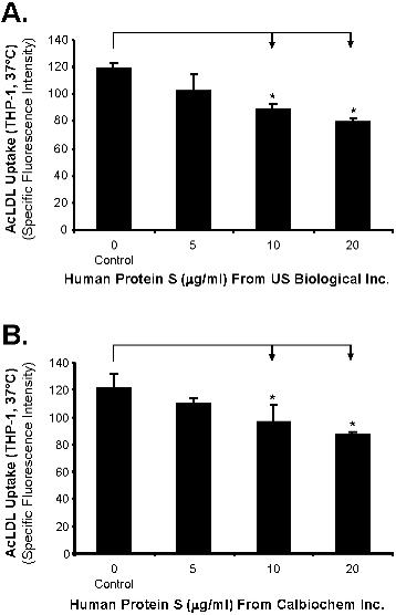
- Figure S2. Effect of protein S from other different sources on Alexa fluor 488-AcLDL uptake in THP-1 macrophages (JPG, 29 KB) -
THP-1 macrophages were treated with human protein S obtained from USBiological Inc. Swampscott, MA (A) or Calbiochem, San Diego, CA (B) for 24 h, followed by incubation with 5 µg/mL Alexa Fluor 488-AcLDL in the presence or absence of 250 µg/mL AcLDL for 3 h at 37°C. The fluorescence intensity of cells was measured by a fluorescence microplate reader.
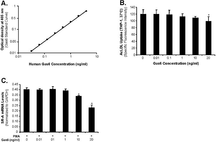
- Figure S3. Gas6 concentration and effects of Gas6 on AcLDL uptake and SR-A mRNA levels in THP-1 macrophages (JPG, 35.3 KB) -
(A) Determination of Gas6 concentratioin. Human Gas6 concentration was determined by using human Gas6 ELISA kit. Human Gas6 concentration standard curve (0.05 to 8 ng/mL) was established. The detection limit of ELISA was 0.03 ng/mL of Gas6. However, Gas6 contamination in commercially-available protein S preparation (1 mg/mL) used in this study was not detected by ELISA. (B) AcLDL uptake. THP-1 macrophages were treated with human Gas6 for 24 h, followed by incubation with 5 µg/mL Alexa Fluor 488-AcLDL in the presence or absence of 250 µg/mL AcLDL for 3 h at 37°C. The fluorescence intensity of cells was measured by a fluorescence microplate reader. (C) SR-A mRNA levels. THP-1 macrophages were treated with human Gas6 for 24 h. SR-A mRNA levels were analyzed by real-time PCR quantification. Results were normalized to the amount of GAPDH mRNA. Values are mean ± SD of three experiments. *P
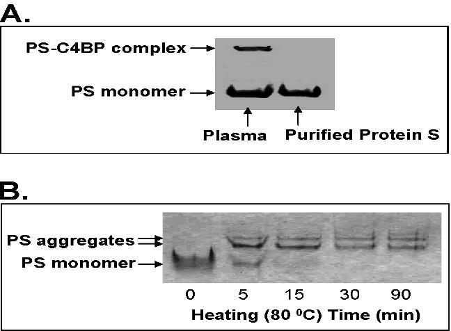
- Figure S4. The detection of non-physiologic protein S aggregation (JPG, 51.6 KB) -
(A) Human protein S (25 ng) or human plasma (1.5 µl) was subjected to native PAGE and detected by western blot analysis. (B) Native gel analysis of protein S. Protein S (2.5 µg) was heated at 80°C for different time points and loaded in 4-12% non-SDS polyacrylamide gel using Tris-glycine buffer. After electrophoresis, the gel was stained with Coomassie blue.
- Figure S5. Effects of human protein S on SR-A expression in primary human aorta smooth muscle cells (JPG, 47.2 KB) -
The primary human aorta smooth muscle cells were treated with 20 µg/mL protein S or 20 ng/mL Gas6 for 24 h. (A) Protein levels of SR-A in primary human aorta smooth muscle cells were detected by western blot analysis. Beta-actin was used as an internal control. Representative results from 3 experiments are shown. (B) SR-A mRNA levels in primary human aorta smooth muscle cells were analyzed by real-time PCR quantification. Results were normalized to the amount of GAPDH mRNA. Values are mean ± SD of three experiments. *P
- Figure S6. The mRNA levels of Axl family members in human macrophages and aorta smooth muscle cells (JPG, 33.9 KB) -
The mRNA levels of Axl, Tyro3, and Mer RTK in U937 monocytes and macrophages (A), human PBMC macrophages (B) and primary human aorta smooth muscle cells (C) were analyzed by real-time PCR. Results were normalized to the amount of GAPDH mRNA. Values are mean ± SD of three experiments.