Blood, Vol. 113, Issue 3, 567-574, January 15, 2009
Foxn1 is required to maintain the postnatal thymic microenvironment in a dosage-sensitive manner
Blood Chen et al. 113: 567
Supplemental materials for: Chen et al
The lacZ allele is indicated as “Z” in all figures.
Files in this Data Supplement:
- Document 1. Supplemental materials and methods (PDF, 64.4 KB)
- Figure S1. Gradual loss of cortical-medullary architecture and reduced thymus size in Foxn1lacZ mutants (JPG, 179 KB) -
(A–F) Hematoxylin and eosin stained paraffin sections of thymi from +/lacZ,lacZlac/Z, lacZ/nu, and +/nu at newborn (A), 1 week (B), 2 weeks (C), 3 weeks (D), and 4 weeks (E). The normal postnatal thymus displays dramatic shifts in size and phenotype over the life of the animal. (A) Newborn +/lacZ and lacZ/lacZ thymi were similar, while thymi from newborn +/nu and lacZ/nu mice were smaller and less well-organized, showing that even a 50% reduction in Foxn1 dosage influenced thymus development. (B) At 1 week, all genotypes showed normal cortico-medullary junction (CMJ) organization. (C) By 2 weeks, lacZ/lacZ and lacZ/nu thymi had a noticeable decrease in cortical area, and lacZ/nu also showed initial CMJ disorganization. (D) CMJ disorganization began at 3 weeks in lacZ/lacZ, while lacZ/nu degeneration progressed further. (E) All phenotypes were progressive at 4 weeks. Scale bar in the upper panel of (A): 1 mm, applies to all upper panels. Scale bar in the lower panel of (A): 100 µm, applies to all lower panels. (F) Thymus weights of different genotypes at newborn and 5 weeks. Newborn +/+ and lacZ/lacZ have similar thymus weight, but thymus weight of +/nu is significantly lower (p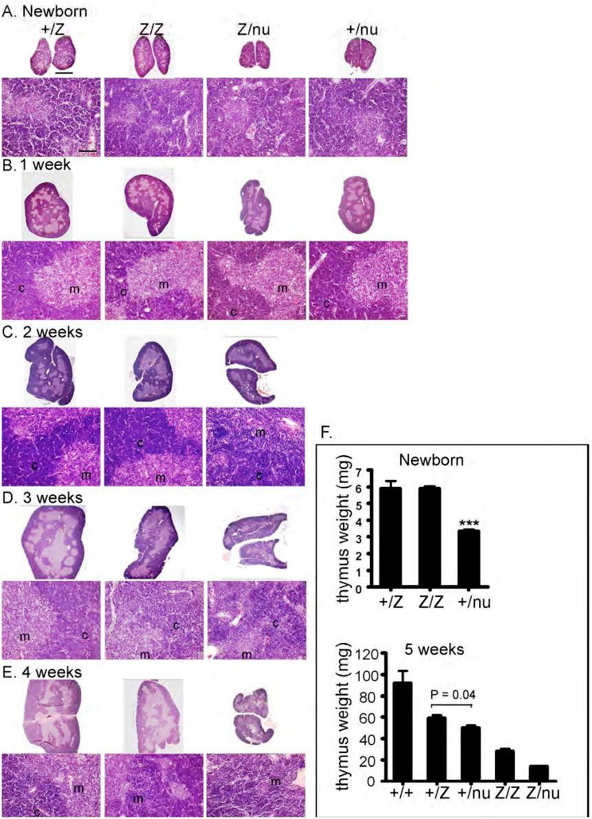
- Figure S2. Primary cultures of thymic stromal cells (TSCs) contain mostly thymic epithelial cells (TECs) (JPG, 59.8 KB) -
(A) Cultured TSCs derived from Foxn1cre/lacZ;R26YFP mice were analyzed for YFP with flow cytometry. More than 70% of the total cultured TSCs are YFP+ cells, which are TEC lineage cells. (B) Most of the cultured TSCs of Foxn1cre/lacZ;R26YFP mice are YFP+. (C) Cultured TSCs stained for the expression of Keratin 14, a marker for medullary TECs. The majority of the cells are K14+.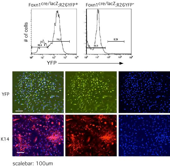
- Figure S3. The epithelial architecture in Foxn1lacZ thymus is relatively normal at newborn and 1 week (JPG, 120 KB) -
Cryosections of +/lacZ, lacZ/lacZ, lacZ/nu, and +/nu thymi at newborn and 1week were stained with anti-Keratin8 (green) and anti-Keratin5 (red) (A) or with anti-Keratin 14 (red) and UEA-1 (green) (B). Small, proto-medullary regions were seen at newborn, and clearly defined organized medullary compartments were formed at 1 week in all four genotypes. Note scattered UEA-1+ cells in newborn +/nu and lacZ/nu thymi due to the haploinsufficiency of the nu allele. Scale bar: 200 µm, applies to all panels.
- Figure S4. Sorting gates of TEC subsets shown in Fig. 2D (JPG, 43.6 KB) -
Gated CD45-thymic stromal cells from 4 weeks old +/+ mice were sorted with flow cytometry for MHCIIhigh versus MHCIImiddle/low (A), and for UEA-1high versus UEA-1middle/low (D). Sorted cells were checked for purity: MHCIImiddle/low (B), MHCIIhigh (C), UEA-1middle/low (E), and UEA-1high (F).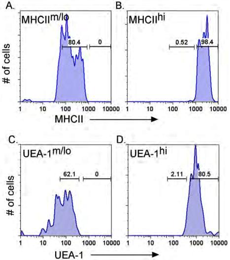
- Figure S5. BrdU analysis of TSC from 1 month old +/lacZ and lacZ/lacZ thymus (JPG, 63.5 KB) -
CD45− cells were gated and stained for UEA-1 and MHCII expression. Total TSC and specific subsets as shown in Fig. 4E were analyzed for the percentage of BrdU+ cells. A summary of these data is shown in Fig. 4F.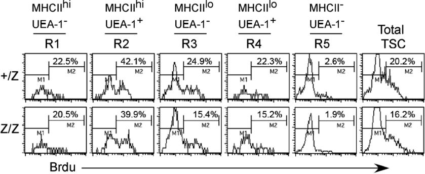
- Figure S6. The hemopoietic cells in Foxn1lacZ mutant are not affected (JPG, 66.3 KB) -
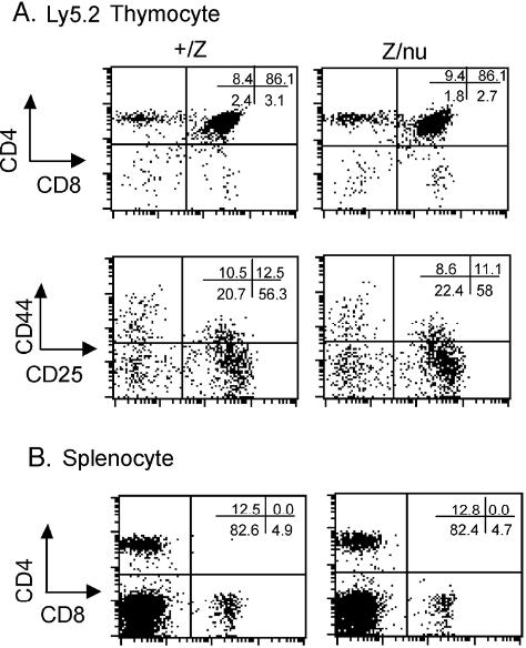
Bone marrow of adult +/lacZ and lacZ/nu mice were transferred into irradiated BL6Ly5.1 mice. (A) Ly5.2+ thymocytes were analyzed 4 weeks after reconstitution for CD4 and CD8 expression, and gated CD4−CD8− cells were analyzed for CD44 and CD25 expression. (B) Splenocytes were also analyzed as controls.