Blood, Vol. 113, Issue 9, 1919-1928, February 26, 2009
Genetic deletion of JAM-C reveals a role in myeloid progenitor generation
Blood Praetor et al. 113: 1919
Supplemental materials for: Praetor et al
Files in this Data Supplement:
- Table S1. Engraftment capacity of JAM-C+ BM cells (PDF, 28.3 KB) -
Recipient mice were considered to be reconstituted by CD45.2+ cells if the frequencies of CD45.2+ donor-derived myeloid (M, CD11b+/Gr-1+) or lymphoid (L, B220+, or CD3ε+) cells detected in the peripheral blood at the indicated times after transplantation were greater than background levels (0.13% CD45.2+, 0.4% B220+, and 0% CD3+ and CD11b/Gr-1+ as determined by analysis of untransplanted BL6/SJL mice). M + L indicates multilineage reconstitution. - Table S2. Hematologic parameters in wt and homozygous mice (PDF, 26.8 KB) -
Peripheral blood samples were collected from wild type (n = 10) and homozygous (n =12) mice at 6–10 weeks of age or from E18.5 wild type (n = 5) and homozygous (n = 4) embryos. Data are mean +/− SE; (ND) not detected. The increase of neutrophils is significant at the p = 0.049 level. - Table S3. Hematopoietic compartments in wt and homozygous mice (PDF, 50 KB) -
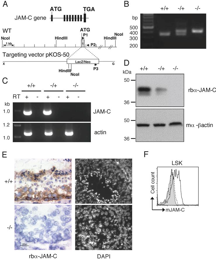
Peripheral blood samples (wt n = 3 and ko n = 4), spleens (wt n = 5 and ko n = 6), thymi (n = 4), BM (wt n = 5 and ko n = 6), or FL (wt n = 11 and ko n = 12) were collected from wild type and homozygous mice. Animals were 4–10 weeks of age and embryos at stage E14.5. Cells were stained for the indicated cell surface markers and analyzed by flow cytometry. Data are mean +/− SE. - Figure S1. Targeted disruption of the JAM-C gene (JPG, 110 KB) -
(A) Schematic diagram of the JAM-C locus including maps of the wild-type (wt) allele and targeting vector, showing the targeted allele (LacZ/Neo: lacZ and neomycin-resistant gene). Black arrows indicate PCR primers (P1, P5, and Neo3a). (B) PCR analysis of genomic tail DNA verifying homologous recombination and presence of neo cassette. PCR product of wild type allele is 347 bp and that of the targeted allele 412 bp. (C) RT-PCR analysis showing JAM-C transcript in testis of wild type, heterozygous, and homozygous mice. Actin is shown as control transcript. (D) Western blot analysis showing expression of JAM-C protein in testis of wild type, heterozygous, and homozygous mice. Actin is shown as loading control. (E) Immunohistochemical staining for JAM-C in testis from wild type and homozygous mice and DAPI stained sections of testicular seminiferous tubules from wild type and homozygous mice, demonstrating spermatid arrest with lack of mature spermatozoa in homozygous seminiferous tubules. Original magnification 60×. (F) JAM-C expression on BM derived LSK cells of wild type (black histogram line) and homozygous mice (hatched histogram line) measured by flow cytometry (LSK: Lin−, Sca-1+, c-kit+). Shown is a representative histograms with rabbit serum IgG (filled grey histogram) as control.