Files in this Data Supplement:
Fig. S1. A representative fragment of the ZBP1 promoter with hypermethylated CpG sites in MTLn3 cells. Red ‘C’ indicates methylated cytosines. Numbered positions are relative to the transcription start site.
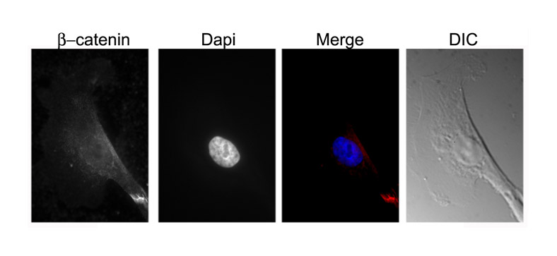
Fig. S2. Immunofluorescence showing the cellular localization of β-catenin in breast non-tumorigenic MCF10 cells (average 100 cells were examined). β-catenin was not localized in cell nuclei (100%). Scale bar: 10 m.
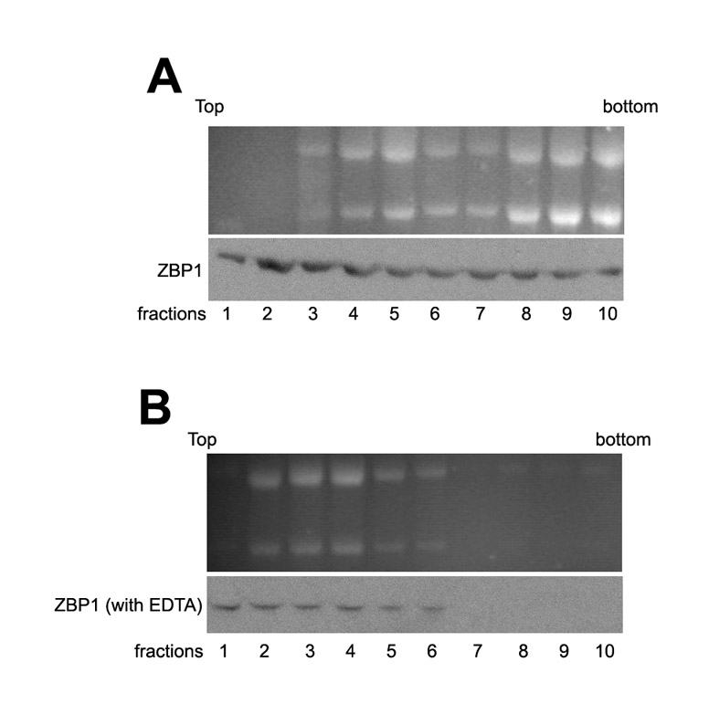
Fig. S3. Extracts of MTLn3 and MTLn3-ZBP1 cells were fractionated in 10-50% linear sucrose gradients in the presence (A) or absence (B) of EDTA. Total RNA was extracted from the fractions and visualized by ethidium-bromide staining after electrophoresis (upper panels, respectively). Western blots show the distribution of ZBP1 from fractions of the sucrose gradients without or with EDTA (lower panels, respectively).