Blood, Vol. 113, Issue 21, 5206-5216, May 21, 2009
A proliferation-inducing ligand mediates follicular lymphoma B-cell proliferation and cyclin D1 expression through phosphatidylinositol 3-kinase–regulated mammalian target of rapamycin activation
Blood Gupta et al. 113: 5206
Supplemental materials for: Gupta et al
Files in this Data Supplement:
- Figure S1. Syndecan expression on FL cells (JPG, 49.2 KB) -
(A) Karpas, DOHH2, FL patient specimens (n=3), and CD19+ B cells from healthy donors (n=3) were stained with PE-conjugated anti-syndecan 1 (thick line) or isotype control (solid gray histogram) and analyzed by flow cytometry.
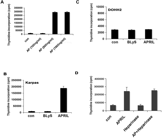
- Figure S2. APRIL-mediated proliferation of FL cells (JPG, 72.6 KB) -
(A) Karpas cells were cultured with increasing concentrations of APRIL for 48 hrs and proliferation was assessed. (B) Karpas or (C) DOHH2 cells were cultured with APRIL (500 ng/ml) or BAFF (100 ng/ml) for 48 hrs and proliferation was assessed. (D) Karpas cells were cultured alone or with APRIL, heparinase, or both for 48 hrs and proliferation was assessed by thymidine incorporation.
- Figure S3. Inhibition of TACI and BCMA in DHL-6 cells (JPG, 81.5 KB) -
Control, TACI, or BCMA siRNA (100 nM) transfected DHL-6 cells were analyzed by flow cytometry and western blot using TACI (A) or BCMA (B) specific antibodies as described in the Material and Methods.
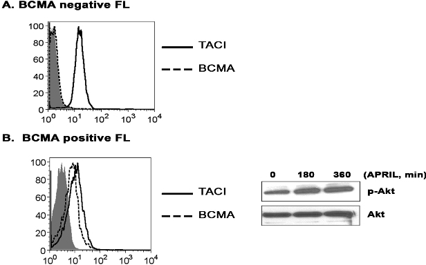
- Figure S4. APRIL-mediated phorphorylation of Akt in BCMA positive and BCMA negative FL patient specimens (JPG, 59.4 KB) -
(A) Expression of BCMA and TACI was analyzed by FACS on the FL patient used for p85 and Akt phosphorylation analysis in Fig. 4F. (B) BCMA positive FL cells, as determined by FACS, were stimulated for the indicated times with APRIL and phosphorylation of Akt (Ser 473) was analyzed by western blot.
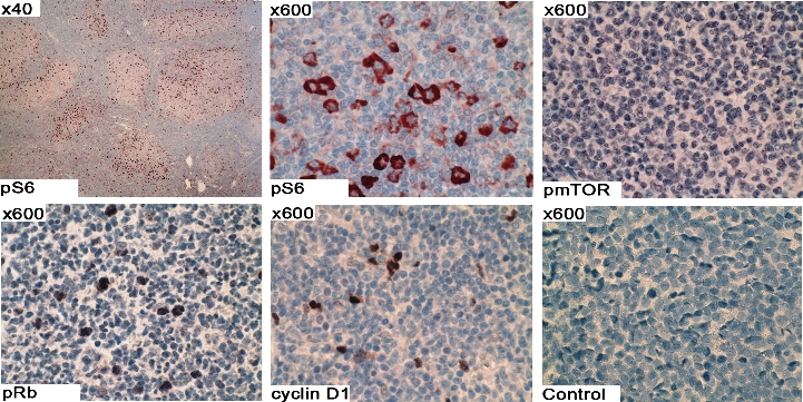
- Figure S5. Activation of the Akt pathway in FL tissue (JPG, 315 KB) -
Phospho-S6 (Ser 235/236) (2211L, Cell Signaling, Danvers MA), phospho-mTOR (ser2448) (NB600–607, Novus Biologicals, Littleton, CO), phospho-Rb (Ser 780) (NB100-92621, Novus Biologicals), and cyclin D1 (RM-9104-S, Thermo Fisher Scientific, Fremont, CA) expression was assessed in FL tissue (n=10) by immunohistochemistry as described in Materials and Methods. An isotype control antibody is shown in the lower right panel. Image magnification is shown in the upper left corner.