Files in this Data Supplement:
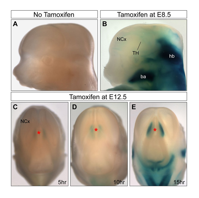
Fig. S1. Temporal response of CreER to tamoxifen administration in Gbx2CreER/+; R26R embryos. (A-E) X-gal staining of Gbx2CreER/+; R26R embryos at E12.5 with and without tamoxifen administration. No β-gal-positive cells were detected in Gbx2CreER/+; R26R embryos without tamoxifen (A). Administration of tamoxifen at E8.5, 48 hours prior to the onset of Gbx2 expression in the thalamus, labels cells in the hindbrain but not in the thalamus (B), indicating that 48 hours after oral gavage administration, tamoxifen fails to activate CreER. β-gal-positive cells in the thalamus of Gbx2CreER/+; R26R embryos at E12.5 are first detected at 5 hours (C), and the number of positive cells progressively increases at 10 hours (D) and 15 hours (E) after a single oral administration of 6 mg tamoxifen. ba, branchial arch; TH, thalamus; ET, epithalamus; hb, hindbrain; mb, midbrain; Ncx, neocortex; PT, pretectum; PTh, prethalamus.
Fig. S2. X-gal staining and BrdU immunohistochemistry of E18.5 Gbx2CreER/+; R26R embryos after low-dose tamoxifen administration at late E9.5 and BrdU injection at E13.5. Distribution of the labeled cells in different regions of the thalamus shows that earlier born thalamic cells, marked by X-gal (blue), have a lateral-to-medial gradient, whereas the later born thalamic cells, marked by BrdU (brown), have a medial-to-lateral gradient, indicating that thalamic cells are deposited in an ‘outside-in’ order according to their time of cytogenesis. However, these two groups of cells are not strictly deposited into separate domains, suggesting that some degree of mixing of cells may exist during development of the thalamus.