Files in this Data Supplement:
Fig. S1. Biochemical quantification of melanin in gart and paics mutants. Micrograms of melanin per fish quantified at 3.5 dpf and 5 dpf. gart and paics mutants possess significantly less melanin at both time points (*** P<0.001).
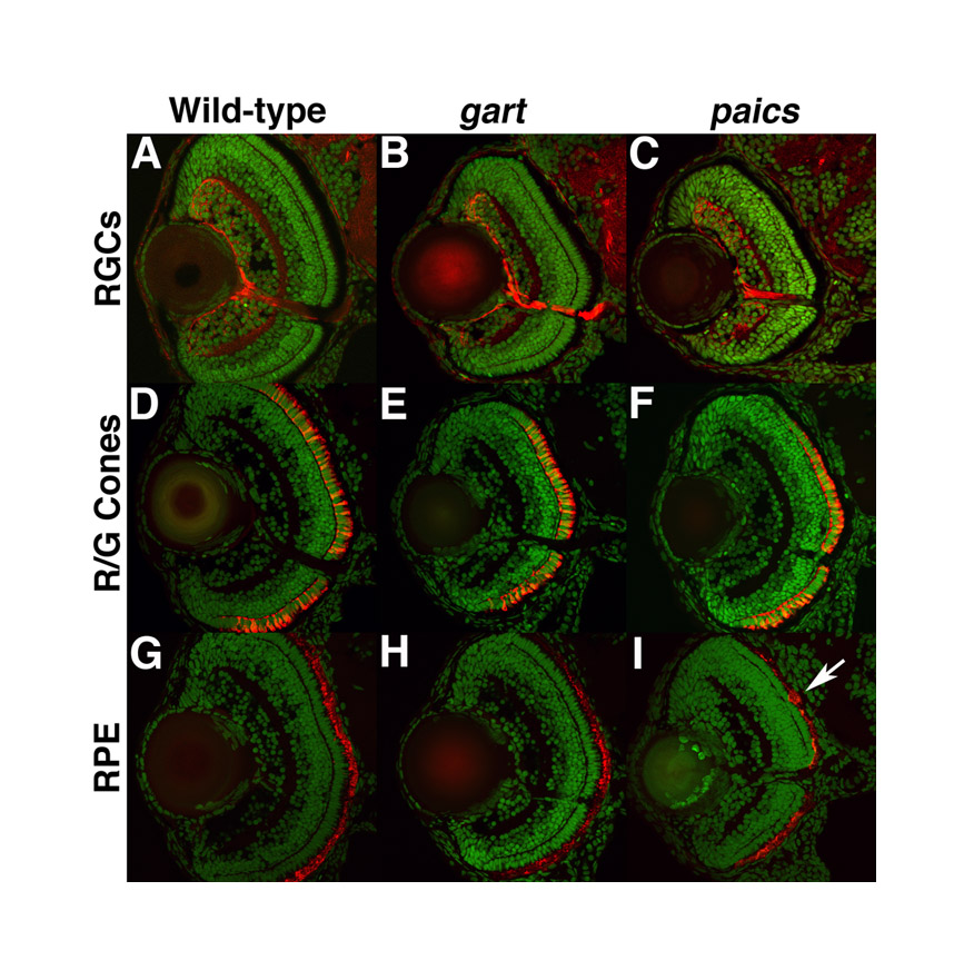
Fig. S2. Immunohistochemical analysis of retinal patterning in gart and paics mutants. Transverse retinal cryosections from 5 dpf embryos stained with zn8, a marker of RGCs (A-C); zpr1, a marker of red/green cones (D-F); and zpr2, a marker of the RPE (G-I). No differences between wild type (A,D,G), gart (B,E,H) or paics (C,F,I) were observed, although occasional clumps of RPE were noted in some mutants (arrow in I).
Fig. S3. A subset of homozygous gart and paics mutants are viable and develop into normal adults. Heterozygous (A) and homozygous (B) gart mutants at 90 dpf. Heterozygous (C) and homozygous (D) paics mutants at 60 dpf. Both gart and paics homozygous mutants develop into normal adult fish and show no overt adult phenotypes.
Fig. S4. adss and gmps morpholino efficacy. (A) RT-PCR for adss using two primer sets that flank the intron 1/exon 2 targeted region of the transcript. Both primer sets do not amplify any transcript from adss-MO-injected embryos at 24 hpf. wnt5 primers are used as a positive control for each cDNA. (B) RT-PCR for gmps using a primer set flanking the exon 2/intron 2 targeted region of the transcript. gmps-MO injection leads to a splicing defect in gmps transcripts at 24 hpf. (C) gmps-MO injection leads to the removal of an 81 bp portion of exon 2 (exon 2 sequence in black, excised portion in red).