Blood, Vol. 114, Issue 7, 1344-1354, August 13, 2009
Interferon β induces mature dendritic cell apoptosis through caspase-11/caspase-3 activation
Blood Yen and Ganea 114: 1344
Supplemental materials for: Yen and Ganea
FACS analysis
DC (1 × 10⁶) were washed in PBS and incubated for 30 min at 4°C with FITC-conjugated anti-CD80, CD86, CD40, or PE-conjugated anti-CD11c (BD Pharmingen). The specificity of the primary Abs was established with appropriate isotype-matched controls. After extensive washing, stained cells were analyzed using a FACSCalibur flow cytometer (BD Biosciences). Results were collected for 10,000 cells and analyzed using CellQuest software from BD Biosciences or FCS Express from De Novo software.
T-cell proliferation assay
For the CFSE assay, purified BALB/c CD4+ T cells (1 × 10⁶ cells/well) were labeled with CFSE (10µM) and cocultured with allogenic B10.A bone-marrow derived DC (2 × 10⁵, 1 × 10⁵, or 5 × 10⁴ cells/well). After 4 days, CD4+ T cells were collected and subjected to CFSE analysis by FACS. For 3H-thymidine incorporation assay, B10.A DC (4 × 10³ cells) treated with IFNβ, or TNF-α+IL-1β+IL-6+PGE2 in the presence or absence of IFNβ for 24h were washed extensively and cultured with purified BALB/c CD4+ T cells (2 × 10⁵ cells). On day 3 of coculture, 3H-thymidine (1µCi per well) was added and incorporation was measured after 16h.
Cytokine ELISA
The cytokine production in DC cultures or DC/T-cell cocultures was determined by ELISA. Supernatants were collected from DC (1 × 10⁶ cells/well) after 48h stimulation and subjected to IL-12 ELISA. DC (1 × 10⁵ cells/well) and T cells (2 × 10⁶ cells/well) were cocultured in 24-well culture plates. Supernatants were harvested after 4 days and subjected to ELISA. The detection limits for IL-12, IL-2, and IFNγ was 62.5 pg/ml.
Files in this Data Supplement:
- Figure S1. Effects of IFNβ on the phenotype of cytokine cocktail-matured DC (JPG, 152 KB) -
DC were treated with TNF-α+IL-1β+IL-6+PGE2 in the presence or absence of IFNβ for 24h. The expression of CD40, CD80, and CD86, was analyzed by FACS. Data are representative of two independent experiments.
- Figure S2. Effects of IFNβ on IL-12p70 production of cytokine cocktail-matured DC (JPG, 106 KB) -
(A) DC were treated with TNF-α+IL-1β+IL-6+PGE2 in the presence or absence of IFNβ for 48h. (B) DC were treated with LPS in the presence or absence of IFNβ for 24h. The supernatants were collected and subjected to IL-12 ELISA. Data are representative of three independent experiments.
- Figure S3. Effects of IFNβ on the capacity of DC to stimulate T-cell proliferation (JPG, 257 KB) -
DC were treated with TNF-α+IL-1β+IL-6+PGE2 in the presence or absence of IFNβ. After 24h, cells were washed extensively and cultured with purified CD4+ T cells, labeled with CFSE, at different ratios (DC:T cells: 1:5; 1:10; 1:20). After 4 days, cells were harvested, and the CSFE profile was assessed by flow cytometry (A). DC treated with IFNβ, or TNF-α+IL-1β+IL-6+PGE2 in the presence or absence of IFNβ for 24h were washed extensively and cultured with purified CD4+ T cells. After 4 days coculture, the proliferation was measured by 3H-thymidine incorporation (B). The supernatants from DC/T cells (1:20) cultures were collected and subjected to IL-2 and IFNγ ELISA (C). Data are representative of three independent experiments for A, and of two independent experiments for B and C.
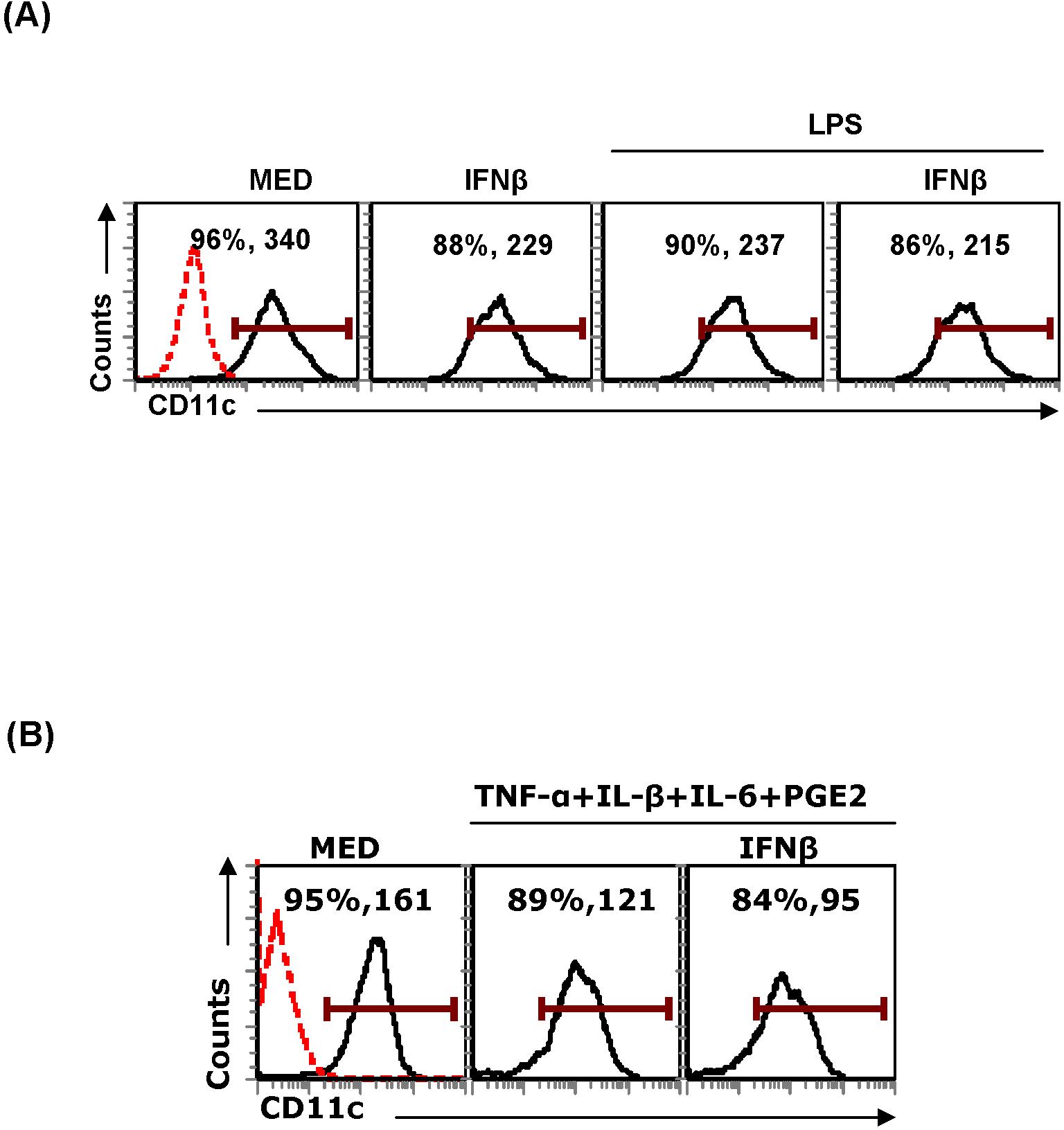
- Figure S4. Effects of LPS and cytokine cocktail on CD11c expression (JPG, 196 KB) -
CD11c+ DC were treated with IFNβ (1000IU), LPS (1µg/ml) in the presence or absence of IFNβ (A) or TNFα+IL-1β+IL-6+PGE2 in the presence or absence of IFNβ (B). After 24h, cells were stained with PE-CD11c followed by FACS analysis. Data are representative of three independent experiments.