Files in this Data Supplement:
Fig. S1. (A) Real-time PCR data showing relative mRNA levels of Coronin 2A and Vinculin in MTLn3 cells expressing shNS, shCoro2A, or Rescue. Note: Rescue is shCoro2A that co-expresses human Coronin2A-EGFP. The real-time probe for Coronin2A does not recognize the human gene. (B) Lysates from 293FT cells expressing human Coronin2A-GFP or rat Coronin2A-GFP were immunoblotted for GFP and with the Coronin 2A antibody. The Coronin 2A antibody recognizes the human protein more strongly than the rat protein. Although we have not quantified the difference in affinity of this antibody for rat versus human Coronin 2A, this observation suggests that the level of re-expression in the rescue cells depicted in Fig. 2A is roughly in the physiological range. This notion is confirmed by the restoration of whole cell motility levels versus the Coronin 2A-depleted cells (Fig. 2B).
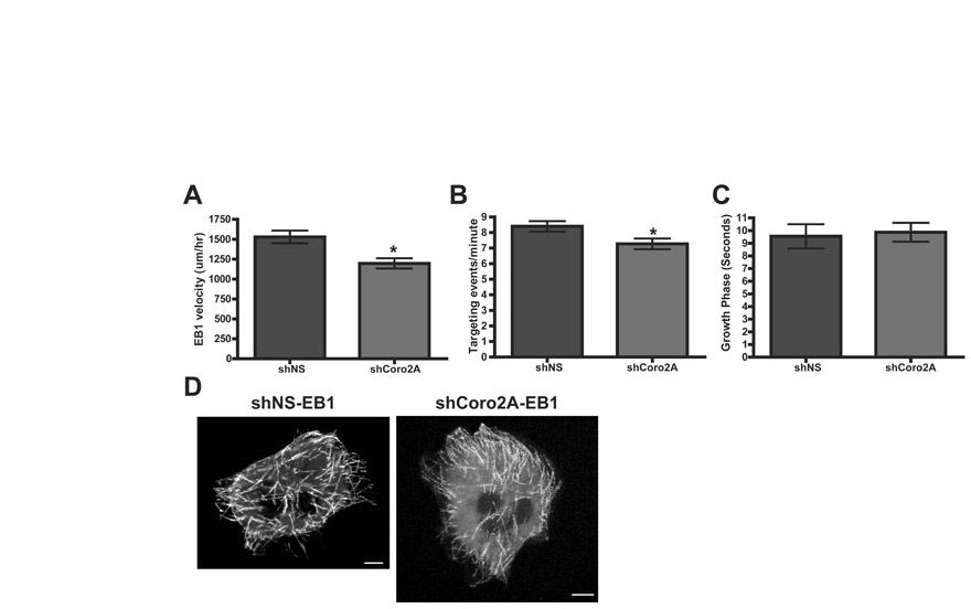
Fig. S2. EB1 dynamics in cells depleted of Coronin 2A. (A) Microtubule growth rate was measured by tracking EB1-GFP velocity. P<0.001. (B) Cells expressing shNS-TRFP-Actin or shCoro2A-TRFP-Actin with EB1-GFP were monitored for occurrences of EB1-GFP in the proximity of the terminal ends of actin stress fibers. This was counted as a targeting event. P<0.001. (C) Growth phase is the amount of time an EB1-GFP spot is visible on a growing microtubule. (D) Example of maximum intensity projections showing EB1-GFP paths. Expression of shCoro2A has no affect on linearity of paths.
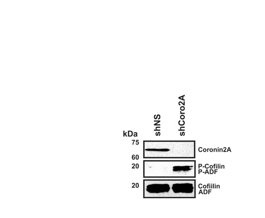
Fig. S3. Depletion of Coronin 2A increases both ADF and Cofilin phosphorylation at Serine 3. MTLn3 cell lysates expressing either shNS or shCoro2A were immunoblotted with an antibody that recognizes both ADF and Cofilin or P-ADF and P-Cofilin. Immunoblot for Coronin 2A shows effective reduction in shCoro2A expressing cells.
Fig. S4. Depletion of Coronin 2A does not affect phalloidin intensity. Cells expressing either shNS or shCoro2A were fixed and stained with Alexa488-phalloidin. Phalloidin intensities were normalized against uninfected neighboring cells. P=0.0572
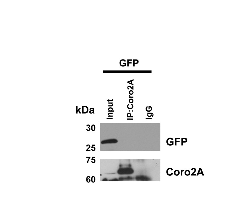
Fig. S5. Coronin 2A does not interact with GFP. 293FT cells expressing GFP were immunoprecipitated for Coronin 2A. Immunoblots of lysates and immunoprecipitations show that Coronin 2A does not interact with GFP.
Movie 1. Whole cell motility of MTLn3 and MTLn3 cells depleted of Coronin 2A (shCoro2A). MTLn3 or shCoro2A cells plated on 50 g/ml rat tail collagen. Images taken every 5 minutes for 4 hours. Left panel shows control cells, and right panel shows shCoro2A cells. Movie speed: 20 frames/second.
Movie 2. Focal adhesion dynamics of control and shCoro2A cells expressing GFP-PXN. Left panel shows control cell, and right panel shows shCoro2A cell. Images taken every minute for 30 minutes.
Movie 3. Live-cell TIRF microscopy movie of MTLn3 cells expressing GFP-PXN and Cofilin-TagRFP. Images taken every minute. Movie speed: 20 frames/second.