Files in this Data Supplement:
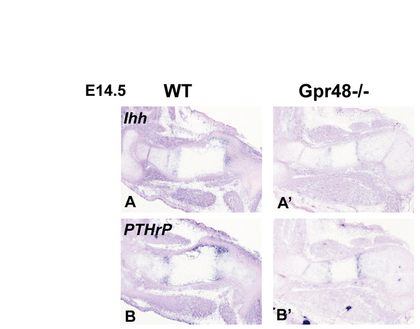
Fig. S1. Deletion of Gpr48 does not disturb Ihh and Pthrp expression. In situ hybridization analysis of chondrocyte differentiation and proliferation markers in wild-type (WT) (A,B) and Gpr48−/− (A′ ,B′ ) femur at E14.5. Probes are Ihh (A,A′) and Pthrp (B,B′).
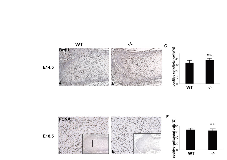
Fig. S2. Deletion of Gpr48 does not disturb chondrocyte proliferation. (A-C) BrdU incorporation in the chondrocyte proliferation zone of E14.5 embryos (A,B). The BrdU-positive cells in the columnar area were counted (brown nuclei). Statistical significance was assessed by one-way analysis of variance and unpaired Student’s t-test. (C) Bar chart showing the percentage of BrdU-positive cells relative to total chondrocyte cell number. n.s., not significant. (D-F) PCNA staining of the chondrocyte proliferation zone of E18.5 embryos (D,E). (F) Bar chart showing the percentage of PCNA-positive cells relative to total chondrocyte cell number.
Fig. S3. Pthrp and Pthr1 expression in primary cultured and differentiating bone marrow mesenchymal cells in vitro. There was no significant difference in Pthrp or Pthr1 expression between wild-type and Gpr48-null mice during osteoblast differentiation (day 0-10).
Fig. S4. Deletion of Gpr48 does not disturb serum Ca2+ or phosphate. There was no significant difference in serum Ca2+ or phosphate between 8- to 16-week-old wild-type and Gpr48-null mice (n=20).