 -tubulin-complex proteins
Files in this Data Supplement:
Fig. S1. Conservation of grip1 motif in A. nidulans and human GCPs. Sequence alignments of A. nidulans and human GCPs. Sequence alignments were generated using the ‘T-coffee’ algorithm. Standard RasMol colors are used for amino acids and the numbers refer to the amino acid number within each protein.
Fig. S2. Conservation of grip2 motif in A. nidulans and human GCPs. Sequence alignments of A. nidulans and human GCPs. Sequence alignments were generated using the ‘T-coffee’ algorithm. Standard RasMol colors are used for amino acids and the numbers refer to the amino acid number within each protein.
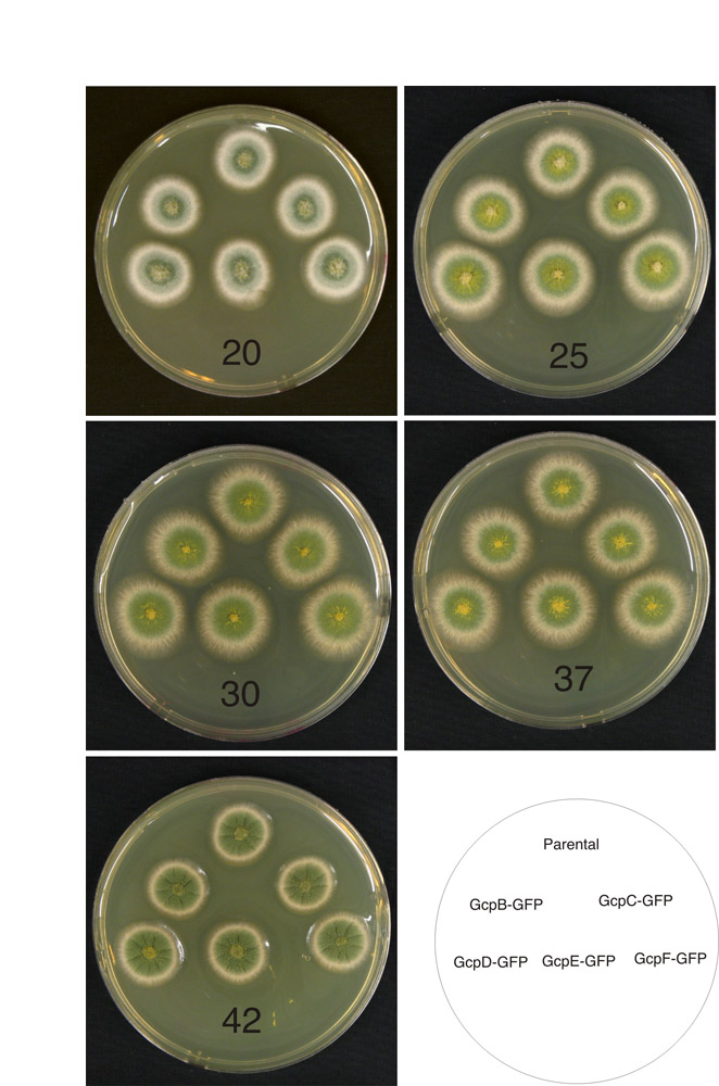
Fig. S3. Growth of strains carrying GCP-GFP fusions. The temperature of incubation is given at the bottom of each plate. Schematic diagram showing the positions of the colonies carrying the various GFP fusions is at the lower right. The GFP fusions do not affect growth rates at any temperature.
Fig. S4. Co-localization of GCPs with the A. nidulans Nud1 homolog. Each row of images shows the same field. Each of the GCPs co-localizes with the A. nidulans Nud1 homolog, a spindle pole body marker. All images are the same magnification.
Fig. S5. Growth of strains carrying deletions of gcp genes. The temperature of incubation is at the upper left of each panel and a schematic showing the genotypes of the strains with respect to the gcp genes is at the upper left. The growth of the gcpΔ strains is indistinguishable from the parental strain at all temperatures.
Fig. S6. Growth and benomyl sensitivites of strains carrying gcp deletions. The left column of plates shows growth of the gcp deletants in a strain expressing GFP-α-tubulin and histone H1-mCherry. The gcp deletant strains grow identically to the parental strain at all temperatures. The right column shows the growth of the same strains on the concentrations of benomyl shown. The gcp deletants are indistinguishable from the parental strain in benomyl sensitivity.
Fig. S7. Recombinant progeny in a cross of two gcpD-F triple deletion strains. Strain LO2018 carrying deletions of gcpD-F and wA3, which confers white conidia, was crossed to strain LO1930, which also carries deletions of gcpD-F but carries fwA1 (fawn conidia). Progeny of the cross were spread on the plate shown. The fact that colonies with fawn, white and green colonies are present indicates that the two strains have crossed successfully. In particular, all colonies with green conidia are recombinant.
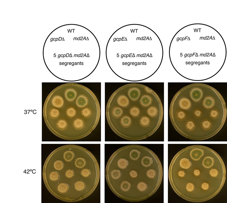
Fig. S8. Synthetic sickness of a deletion of the A. nidulans MAD2 homolog (md2AΔ) and deletions of γ-tubulin complex proteins. A schematic diagram of the genotypes of the colonies is shown over each pair of plates and the temperature of incubation is shown at the left. md2AΔ and gcpD and gcpE deletions show weak synthetic interactions. Colonies carrying the double mutants are slightly smaller than either parent and have rough edges at 37°C. The md2AΔ, gcpFΔ interaction is strongest.
Fig. S9. A matrix surrounds the ends of microtubules at the SPB. The four panels are from consecutive serial sections through a SPB in mitosis. Section A is in the cytoplasm, B in the SPB (arrow), C in the inner SPB and D in the nucleus. Microtubules are visible in C as circular outlines in the SPB matrix. Specimen preparation and observation procedures were as previously described (Oakley and Morris, 1983).