Files in this Data Supplement:
Fig. S1. pigs is expressed in the somatic tissues of the ovary. (A-C) The Gal4 P-element trap inserted into the second intron of pigs was used to reveal pigs expression in the ovaries. Intron2-Gal4 was crossed to the UAS-GFP-EFGas2 transgene (Maybeck and Röper, 2009). Expression is detected in the terminal filament and cap cells (A), the escort cells (B, arrows) and the stalks (C).
Fig. S2. Additional cell shape and cytoskeletal phenotypes observed in pigs1 mutant ovarioles. (A,B) The normal hexagonal packing pattern of follicle cells (FCs) found in wild-type egg chambers (A, cell outlines labelled by HtsF) is disrupted in pigs1 mutant egg chambers and FCs show irregular shapes (B, labelling is Shot). (C-E) Ring canal morphology is aberrant in pigs1 mutant egg chambers (in 30.8% of ovarioles; rescue improves this to 19%). Wild-type ring canals are smooth and round (inset in C) and ring canals in pigs1 mutants often show irregular morphology (C,E). The ring canal component HtsF is properly localised to the inner rim in the pigs1 mutant (D). (F) Many stage 10B egg chambers are dumpless in pigs1 mutants. (G-K) During dumping, nurse cell nuclei are held in place by a �cage� of actin filaments (G, wild-type). pigs1 mutant egg chambers show disrupted actin cages that appear to only form on one side of the nucleus and fail to anchor to the nurse cell membrane (H,J). Nurse cell nuclei can be found blocking the ring canals (I). (K,L) The cap cells of the germarial niche show strong cortical actin staining in the wild-type (K, bracket). This actin enrichment is unchanged in pigs1 mutant germaria (L, bracket). (M) Ovariole with a fully pigs1 mutant follicle cell epithelium but wild-type germline (mutant cells are marked by absence of GFP, green). In these complete FC clones, stalks were malformed or missing, egg chamber encapsulation failed and FCs showed aberrant migration behaviour as in the pigs1 mutant. GFP is in green, nuclei/DAPI in blue and actin/Phalloidin in red.
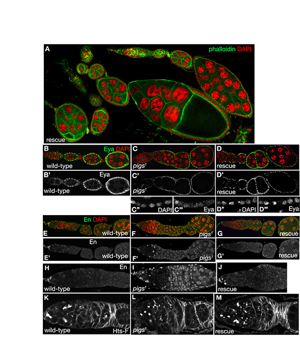
Fig. S3. Rescue of pigs1 phenotypes.pWR-Ubi-pigs cDNA was expressed in the pigs1 mutant background (termed �rescue�) and recovery of phenotypes was quantified in comparison to wild-type and pigs1 mutant ovarioles as detailed in Table 1. This figure shows representative immunofluorescence stainings of the rescue. (A) Overview of ovarian morphology in the rescue, with ovarioles showing individual egg chambers separated by stalks and germaria that bud off egg chambers. (B-D′′′) Rescue of the Eya expression phenotype (Eya in green in B-D and as a single channel in B′-D′,C′′′,D′′′). (E-J) Rescue of the En expression phenotype (En in green in E-G and as a single channel in E′-G′,H-J). H-J show higher magnification of E-G. (K-M) Rescue of the excess GSC number phenotype. Germaria are labelled for Shot.
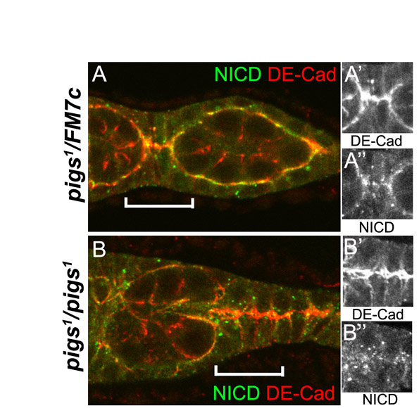
Fig. S4. Pigs affects NICD localisation. (A-B′′) The Notch intracellular domain (NICD) is mislocalised in pigs1 mutant ovarioles, indicating that Pigs could function to localise Notch correctly. (A-A′′) In the wild-type, in those FCs in region 2-3 of the germarium that are ingressing to interdigitate and form the pre-polar and pre-stalk cells, E-Cadherin is strongly localised at the cell-cell contacts (red and single channel in A′). The NICD also mostly localises to these contact zones and the cell surface (green and single channel in A′′). (B-B′′) In pigs1 mutant ovarioles, the early FCs fail to interdigitate, sometimes resulting in long parallel rows of cells. The NICD (green and single channel in B′′) is not concentrated where E-cadherin is (red and single channel in B′) but it appears punctate and intracellular.