Blood, Vol. 115, Issue 19, 3869-3878, May 13, 2010
Therapy of relapsed leukemia after allogeneic hematopoietic cell transplantation with T cells specific for minor histocompatibility antigens
Blood Warren et al. 115: 3869
Supplemental materials for: Warren et al
Files in this Data Supplement:
- Table S1. Primer sequences for RT-PCR analysis of expression of mHAg-encoding genes (PDF, 33.4 KB)
- Table S2. SNPs showing potential association with the mHAg recognized by CTL clone 11C6-109 (PDF, 49.3 KB)
- Table S3. Correlation between genotype at rs7958311 and recognition of HLA-2902+ EBV-LCL by CTL clone 11C6-109 (PDF, 40.1 KB)
- Table S4. SNPs showing potential association with the mHAg recognized by CTL clone 50F5-448 (PDF, 49.3 KB)
- Table S5. Correlation between genotype at rs35394823 and recognition of HLA-5701+ EBV-LCL by CTL clone 50F5-448 (PDF, 38 KB)
- Figure S1. Representative CD8+ CTL clone specific for a tissue-restricted mHAg and used in adoptive T-cell therapy of relapse (JPG, 100 KB) -
Cytolytic activity of CD8+ CTL clone 11C6-109, administered to Patient #1, against recipient- and donor-derived EBV-LCL and dermal fibroblasts, as well as the recipient’s ALL blasts obtained at the time of initial posttransplant relapse, prior to T-cell therapy.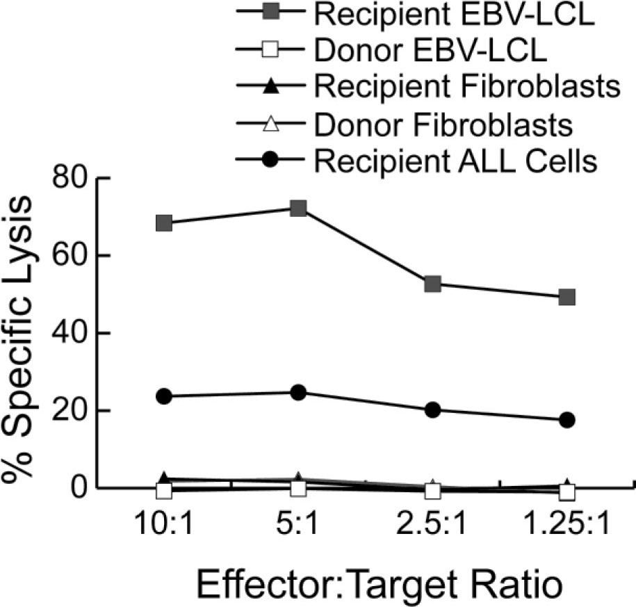
- Figure S2. The P2RX7265–273 peptide WFHHCHPHY is the mHAg recognized by CD8+ clone 11C6-109 (JPG, 130 KB) -
(A) Recognition by 11C6-109 CTL of COS-7 cells that were transiently transfected with plasmid vectors encoding the indicated peptide sequences in the presence (black columns) or absence (light columns) of a plasmid encoding HLA-A29, as determined by IFN- ELISA. (B) Recognition of donor-derived, mHAg-negative EBV-LCL pulsed with the indicated concentrations of the P2RX7265–273 peptide WFHHCHPHY or the homologous peptide WFHHCRPHY encoded by the antigenic and nonantigenic alleles of P2RX7, respectively, in a four-hour 51Cr cytotoxicity assay at E:T 5:1.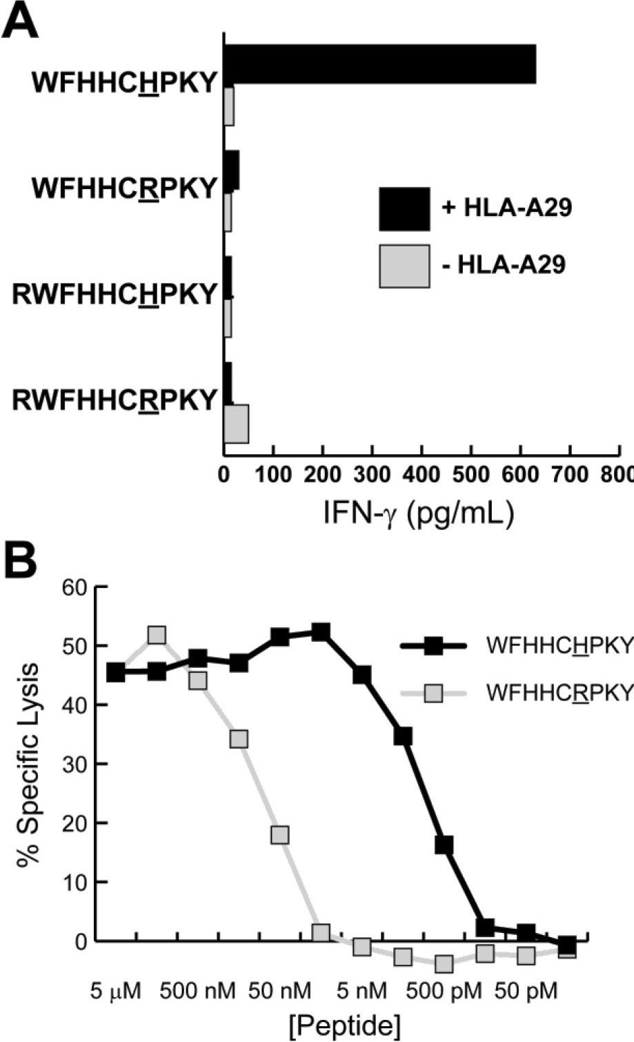
- Figure S3. Consistent expression of MHC class I and adhesion molecules in leukemic blasts from Patient #1 before and after T-cell therapy (JPG, 207 KB) -
PBMC containing >90% leukemic blasts obtained from Patient #1 at the time of the first (blue lines) or second (red lines) relapse were analyzed by flow cytometry for expression of MHC class I, HLA-A29, CD54, CD58, CD44, and PD-L1. Cells stained with an isotype control mAb are indicated by the green lines.
- Figure S4. The DPH1334–343 peptide SVLPEVDVW is the mHAg recognized by CD8+ clone 50F5-448 (JPG, 124 KB) -
(A) Recognition by 50F5-448 CTL of COS-7 cells that were transiently transfected with plasmid vectors encoding the indicated peptide sequences in the presence (black columns) or absence (light columns) of a plasmid encoding HLA-B57. (B) Recognition of donor-derived, mHAg-negative EBV-LCL pulsed with the indicated concentrations of the DPH1334–343 peptide SVLPEVDVW encoded by the antigenic allele of DPH1 in a four-hour 51Cr cytotoxicity assay at E:T 5:1.
- Figure S5. Poor T-cell recognition of leukemic blasts from Patient #7 is not explained by loss of expression of MHC class I or adhesion molecules (JPG, 132 KB) -
PBMC containing >90% leukemic blasts obtained from Patient #7 at the time of posttransplant relapse were analyzed by flow cytometry for expression of MHC class I, CD54, CD58, and CD44 (red lines). Cells stained with an isotype control mAb are indicated by the green lines.