Blood, Vol. 115, Issue 23, 4934-4943, June 10, 2010
Hematopoietic progenitor cell regulation by CD4+CD25+ T cells
Blood Urbieta et al. 115: 4934
Supplemental materials for: Urbieta et al
Files in this Data Supplement:
- Figure S1. CD4+CD25+ T-cell selection and suppressive activity in lymphoid and hematopoietic functional assays (JPG, 178 KB) -
(A) Representative B6-CD8−∕− CD4+CD25+ preparation. CD4+CD25+ T cells (Treg) were prepared from spleen and LN cells by depleting B cells and CD8 T cells (unless CD8−∕−) on Ab coated plates prior to positive selection with anti-CD25 mAb followed by magnetic bead isolation using Miltenyi columns. CD4+CD25+ enrichment was routinely >93% of total cells and >97% CD4+ T cells. Permeabilization of cells and FoxP3 staining was performed using FoxP3-FITC mAb (Clone, FJK-16S, eBioscience, San Diego, CA). (B) Activated B6-CD8−∕− Treg cells inhibit CD4+CD25− stimulated responding syngeneic T cells (see Methods). (C) Activated Tregs can inhibit allogeneic as well as syngeneic marrow. Microbeads and rIL-2 (Methods) were used to activate CD4+CD25+ T cells prepared from B6-CD8−∕− spleen and lymph node cells. Following activation, cells were co-cultured for 72 hrs. with syngeneic (B6) or allogeneic (BALB/c) T-cell–depleted (TCD) BMC. Significant inhibition (p+CD25+ Treg co-cultured with 2.0 × 10⁴ 95% Lin− syngeneic BM for 1.5 days. Analysis of overall number of colonies present at 11 days in cultures supplemented with a “GEMM” cytokine cocktail containing SCF, rmIL-6, rmIL-3, Epo, and TPO (see methods) is presented.
- Figure S2. Treg cell inhibition of CFU-IL3 activity requires MHC class II expression by bone marrow cells and inhibition is detected with lineage negative BMC (JPG, 149 KB) -
(A) Freshly isolated CD4+CD25+ T cells were prepared from B6-CD8−∕− spleen cells and activated as described (Methods). Following 72 hrs., either TCD-BMC from B6-wt or B6-MHC class II−∕− mice were added and the cultures were supplemented with SCF and rmIL-3 as described (Methods). Following 48 hrs., cultures were harvested and plated in methylcellulose for CFU detection. Inhibition was observed in cultures containing Treg cells plus B6-wt but not B6-MHC class II−∕− TCD-BMC. (B) Independent experiment in which following activation, Treg cultures from B6-CD8−∕− mice were washed and lineage depleted, c-kit enriched BMC from B6-WT or B6-MHC class II−∕− mice were added. Lineage depletion was performed by treatment with an ab cocktail (Lineage depletion kit, Miltenyi Biotech, Auburn, CA 95602) followed by negative selection via miltenyi column passage. C-kit enrichment was performed using anti–c-kit–PE (BD Biosciences, San Diego, CA) mab and then positive selection following column passage (sse Methods). Lin−ckit+ enrichment, 79%, B6-wt; 92%, B6-MHC class II−∕−. Inhibition was observed only in co-cultures containing B6-wt marrow cells. (C) B6-wt BMC were lineage depleted by fluorescence activated cell sorting (FACSAria, BD) using a mAb cocktail (anti-CD4, CD8, Ter119, B220, Gr-1, Mac-1). Lineage depletion was >99.6%. These cells (3500/well) were mixed with day 3 activated B6-CD8−∕− Tregs (1:2) and cultured for CFU-IL3 activity. Significant inhibition of CFU-IL3 was observed (p+CD25+ T cells from B6-cdd (cytotoxic double deficient, B6-perf−∕−faslgld∕gld) spleen and lymph node cells were co-cultured 3 days with B6-WT TCD-BMC supplemented with SCF and rmL-3 as described in Methods. These cytolytic defective Tregs mediated inhibition of CFU-IL3 in the cultures.
- Figure S3 (JPG, 162 KB) -
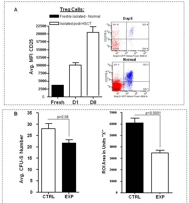
(A) B6 mice (9.5Gy) were transplanted with syngeneic marrow plus B6-FoxP3gfp KI Treg cells. CD25 expression was assessed on splenic and lymph node FoxP3+ CD4 T cells at indicated times post-transplant. Fresh refers to CD25 assessment from pooled (n=5) B6-FoxP3gfp KI splenic and lymph node Treg cells from normal mice. Dot plots represent staining results from an individual B6 mouse transplanted with syngeneic marrow plus B6-FoxP3gfp KI Treg cells 8 days previously or from a normal B6 mouse. (B) B6 mice were conditioned with 9.5Gy TBI and transplanted with 0.15 × 10⁶ TCD-syngeneic BM at Day 0 with (“experimental”) or without (“control”) 1.2 × 10⁶ CD4+CD25+ Treg cells (n=4 mice/group). CFU-S scoring was performed at Day 10 using a dissecting stereoscope (Left Panel). Colony area measurement was performed (right panel) using ImageJ software (NIH). ROI = region of interest.
- Figure S4 (JPG, 155 KB) -
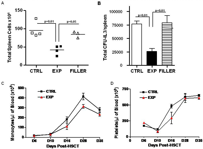
B6 bone marrow (5 × 10⁶ TCD) was transplanted alone (“Control”), or together with 1 × 10⁶ purified B6 CD4+CD25+ Treg cells (“Expt”) or B6 CD19+ B cells (>85%, miltenyi bead positive selection) i.e. “Filler” into 9.5Gy TBI MHC identical recipients (n=3–4/group) (A) Total number of spleen cells 7 days post-transplant. (B) The number of CFU-IL3 per spleen is presented as the average number calculated from each mouse in the transplanted groups following in vitro CFU assay as described (Materials and Methods). Monocyte (C) and platelet (D) were obtained from individual transplant recipients (n=4/group). B6-wt mice (10–12 wk old) were lethally irradiated (9.5Gy TBI) and transplanted with 0.3 × 10⁶ syngeneic TCD-BM. Animals were untreated or co-infused with 1 × 10⁶in vivo IL2/anti-IL2 mAb complex expanded (see Methods) Treg cells (>95% CD4+CD25+) at the time of transplant. Peripheral blood was obtained from animals at indicated days post-HSCTand 40ul were analyzed by the Dept of Pathology using a Hemavet 950.