Files in this Data Supplement:
Fig. S1. olis469 mutants exhibit liver-specific cell death. (A,B) Confocal images of transverse sections of 4-dpf Tg(fabp10:RFP)gz12 WT (A) and olis469 mutant (B) livers stained for cadherin (blue) and TUNEL (green). Although massive cell death occurs in olis469 mutant livers (B), only a few apoptotic cells can be detected in other tissues, such as the gut. The low number of apoptotic cells detected in olis469 mutant tissues other than the liver is equivalent to that found in WT siblings. Bars, 20 µm.
Fig. S2. Mutation in tomm22 in olis469 mutants. (A) Genetic map of the tomm22 region on linkage group 12; the numbers below the SSLP markers indicate the number of recombinants per meioses analyzed (1363 or 1222). The critical region appears to contain eight genes: (1) KIAA1093, (2) casein kinase 1e, (3) MAP kinase activating protein, (4) maff, (5) tomm22, (6) dmc1, (7) gspt1 and (8) ribosomal L1 domain containing 1 (http://vega.sanger.ac.uk/Danio_rerio/Location/View?r=12:14827564-15047873). (B) Sequence analysis of WT (top) and olis469 mutant (bottom) cDNA. A nucleotide change in the tomm22 sequence was identified in olis469 mutants, from thymidine (T) in the WT sequence to cytosine (C). (C) Protein sequence of WT Tomm22 (top) and the predicted Tomm22s469 protein (below). The nucleotide change identified in olis469 mutants results in the substitution of the stop codon (TGA) in WT Tomm22 with an arginine residue (CGA) in the Tomm22s469 protein, leading to a predicted mutant protein with an additional 47 amino acids (aa).
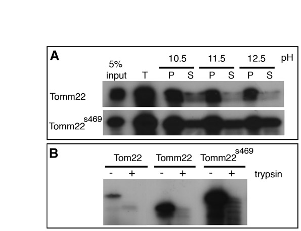
Fig. S3. Tomm22s469 is correctly localized to the yeast outer mitochondrial membrane. (A,B) Radiolabeled WT or mutant Tomm22 was imported into purified yeast mitochondria and then carbonate extracted (A) or trypsin digested (B). (A) Total (T), membrane (P) and soluble (S) fractions are shown and a 5% input lane of in vitro translated product is included as a control. (B) Imports with (+) and without (−) trypsin are shown. Tomm22s469 associates with the membrane fraction after import into yeast mitochondria (A), and both WT and mutant Tomm22 are correctly localized to the yeast outer mitochondrial membrane, as is observed with yeast Tom22 (B).
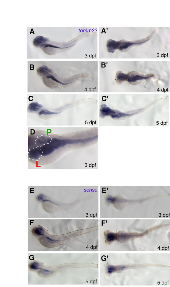
Fig. S4. tomm22 is expressed highly in the endoderm. (A-D) tomm22 expression in WT larvae at 3 dpf (A,A�,D), 4 dpf (B,B�) and 5 dpf (C,C�). A, B and C are lateral views; A�, B�, C� and D are dorsal views. (E-G) In situ hybridization of WT larvae using the tomm22 sense probe as a negative control, at 3 dpf (E,E�), 4 dpf (F,F�) and 5 dpf (G,G�). E, F and G are lateral views; E�, F� and G� are dorsal views. (D) The liver (L) and pancreas (P) are outlined. (A-G�) At 3-5 dpf, tomm22 expression is heightened in the liver, gut and, although not as strongly, the pancreas. Endodermal organ staining was clearly absent when using the sense probe. tomm22 expression could also be detected in the head, although similar, weaker nonspecific staining could also be detected in the head of larvae hybridized with the sense probe.
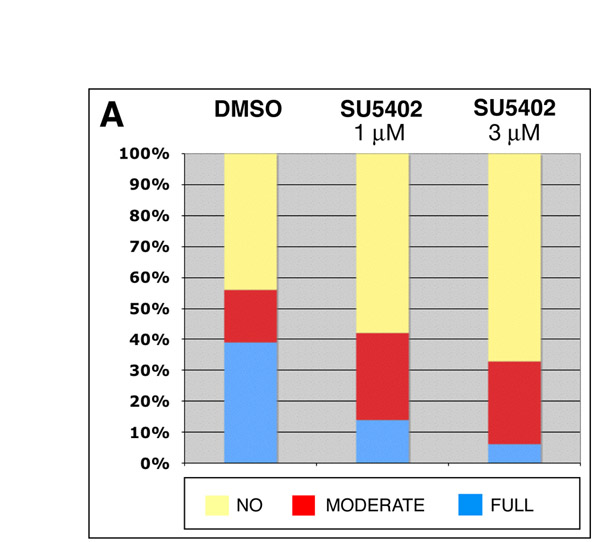
Fig. S5. Role of Fgf signaling in the tomm22 MO hepatocyte regeneration model. (A) Comparison of hepatic recovery post-tomm22 MO-mediated ablation in larvae that were allowed to recover in carrier solution (DMSO; left bar, n=52) versus in the presence of 1 µM (middle bar, n=57) or 3 µM (right bar, n=64) of the FgfR inhibitor SU5402. tomm22 MO-injected larvae that showed severe hepatic ablation at 4 dpf were allowed to recover, and at 6 dpf their liver recovery was classified as (1) �no� change in liver size, (2) �moderate� recovery of hepatic volume, or (3) �full� recovery, comparable to that observed at 6 dpf in most WT larvae injected with 4 ng of tomm22 MO. Compared with the DMSO control larvae, we observed an inhibitory effect on hepatic regeneration, post-tomm22 MO-induced ablation, in the presence of 1 µM SU5402. This inhibitory effect was even more pronounced in the presence of 3 µM SU5402. �Full� recovery (blue) decreased from 39% in DMSO to 14% in 1 µM SU5402 and to 6% in 3 µM SU5402. Conversely, �no� recovery (yellow) increased from 44% in DMSO to 58% in 1 µM SU5402 and to 67% in 3 µM SU5402. �Moderate� recovery (red) was 17% in DMSO, 28% in 1 µM SU5402 and 27% in 3 µM SU5402.