Files in this Data Supplement:
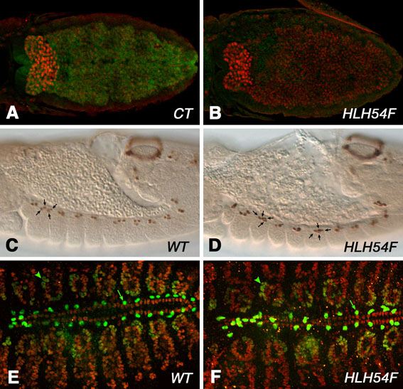
Fig. S1. Mesodermal markers that are not affected in HLH54F mutant embryos. (A) Stage 10 HLH54FS1750/CyO, twi>EGFP control (CT) embryo, stained for Zfh1 and EGFP (dorsal view). Focal plains showing the internal layer with high levels of Zfh1 have been merged. (B) Homozygous HLH54FS1750 mutant embryo, stage and staining as in A. The domain of high Zfh1 expression in the caudal mesoderm is present, although it may be slightly reduced in size. (C,D) Late stage 12 wild-type and HLH54FΔ598 mutant embryos, respectively, showing normal Even-skipped (Eve) expression in 3-4 cells per hemisegment, which correspond to pericardial and somatic muscle 1 progenitors (arrows). (E,F) Stage 16 wild-type and HLH54FΔ598 mutant embryos, respectively, showing normal Eve-stained somatic muscle 1 (arrowheads) and pericardial cell nuclei (arrows), as well as normally arranged nuclei of cardioblasts detected by anti-Mef2.
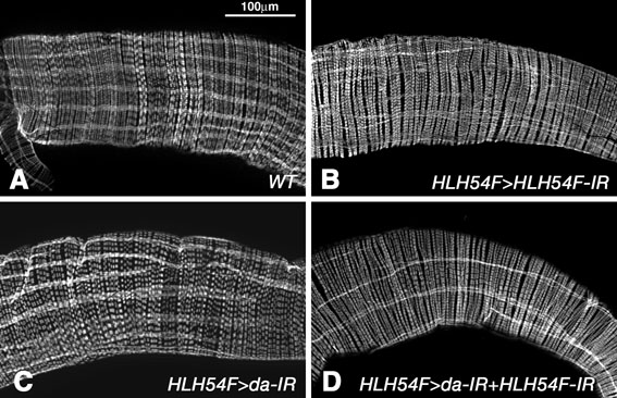
Fig. S2. Effects of RNAi-mediated knockdown of HLH54F and da in adult midgut muscles. The effects of HLH54F-GAL4-driven UAS-HLH54F-IR are significantly delayed as compared with those seen with HLH54F mutations and, like those with UAS-da-IR, are only seen in post-embryonic stages. Shown are adult midguts stained with phalloidin for β3-tubulin (merged). (A) In the wild type, longitudinal gut muscles are spaced evenly around the midgut circumference and display similar widths. (B) Knockdown of HLH54F in longitudinal gut muscles causes a reduction in numbers, thinning, uneven spacing and branching of the fibers. (C) Knockdown of da causes reduction in numbers, uneven spacing and branching of longitudinal gut muscle fibers. (D) Example of knockdown of both HLH54F and da with more severely reduced, branched and aberrantly spaced longitudinal gut muscle fibers.
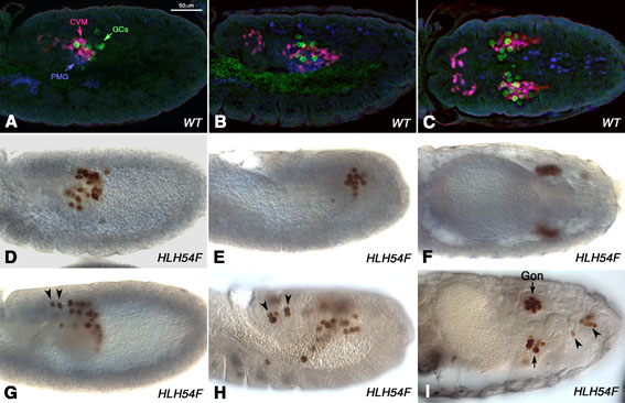
Fig. S3. The role of the CVM in germ cell migration. (A-C) Wild-type embryos carrying HLH54Fb-lacZ and stained for β-gal (CVM, red), Vasa (germ cells, green) and Hb9 (posterior midgut endoderm, blue). (D-I) HLH54F mutant embryos stained for Vasa (brown). (A) Stage 10 embryo prior to CVM migration where germ cells migrating from PMG to trunk mesoderm are in contact with CVM. (B,C) Lateral view (B) and dorsal view (C) of late stage 11 embryo showing close associations of CVM and germ cells during early migration. (D-F) Stage 11, 12 and 14 HLH54FS1750/HLH54FS0323 mutant embryos showing normal germ cell migration and gonad formation. (G) Stage 10 HLH54FS1750/HLH54FS0320 mutant embryo with germ cells having mis-migrated posteriorly (arrowheads). (H) Stage 12 homozygous HLH54FS1750 embryo with some germ cells remaining at the caudal tip (arrowheads) and others near posterior midgut. (I) Stage 15 homozygous HLH54FS1750 embryo with germ cells stuck caudally and gonad(s) containing fewer germ cells.
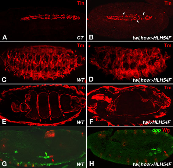
Fig. S4. Effects of ectopic HLH54F expression on mesodermal tissue development. (A,B) Tin-stained dorsal vessels in stage 16 control embryo (CT) and 2xPE-twi-GAL4/+; 24B-GAL4/UAS-HLH54F embryo with pan-mesodermal HLH54F expression, respectively. Ectopic HLH54F causes only a mild reduction of cardioblast formation, which leads to some interruptions in the cardiac tube (arrowheads). (C,D) Stage 16 WT and 2xPE-twi-GAL4/+; 24B-GAL4/UAS-HLH54F embryos, respectively, stained for Tropomyosin. Ectopic expression of HLH54F causes severe morphological defects in the somatic musculature (D). (E,F) Stage 15 embryos with genotypes and stainings as in C,D, but focused on the midgut. Whereas in the wild type there are three midgut constrictions at this stage (E, arrows), none of the midgut constrictions are formed upon ectopic mesodermal expression of HLH54F (F). (G,H) Late stage 11 embryos with genotypes as in C,D, respectively, and stained for dpp mRNA and Wg protein. Whereas wild-type embryos display adjacent dpp (green arrow) and Wg (red arrow) expression domains in the founder cells of parasegment 7/8 of the trunk visceral mesoderm, ectopic expression of HLH54F leads to the loss of the corresponding Wg domain (red arrow) and a broadening of dpp into fusion-competent cells of the visceral mesoderm (green arrow).