Files in this Data Supplement:
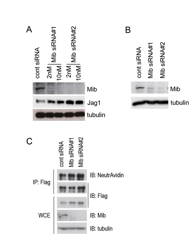
Fig. S1. The expression of Mib is efficiently knocked down by two independent siRNAs, but Jagged 1 expression is not affected by the Mib knockdown. (A) Jag1-3T3 cells were transfected with two Mib siRNAs (#1 and #2) at 2 nM or 10 nM, and whole-cell extracts were immunoblotted with anti-Mib (Mib, upper panel), anti-Flag (Jag1, middle panel), and anti-α tubulin (tubulin, bottom panel) antibodies. (B) Notch-3T3 cells were transfected with two Mib siRNAs (#1 and #2) at 10 nM, and whole-cell extracts were immunoblotted with anti-Mib (Mib, upper panel) and anti-α tubulin (tubulin, lower panel) antibodies. (C) The surface proteins of Jag1-3T3 cells were labelled with Sulfo-NHS-LC-Biotin for 15 minutes, and whole-cell lysates were prepared. Flag-tagged Jag1 was immunoprecipitated with a Flag antibody from the whole-cell extract (WCE). Immunoprecipitates were immunoblotted with NeutrAvidin and the anti-Flag antibody. WCE was immunoblotted with anti-Flag, anti-Mib, and anti-α tubulin antibodies. α tubulin was used as a loading control. IP, immunoprecipitation; IB, immunoblot.
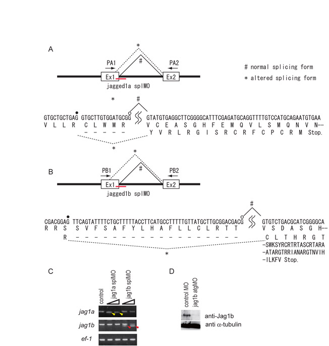
Fig. S2. Morpholino-mediated knockdown of the Jagged genes in zebrafish. (A-C) The jag1a splMO (A) and jag1b splMO (B) were designed to target the splice donor sites of the first exon-intron boundary. These splMOs caused deletions of the mRNAs by altering the splicing donor sites (*; confirmed by DNA sequence analysis of the PCR products indicated by yellow and red arrowheads in C). The altered splicing forms of mRNA potentially produce premature truncated proteins (A,B). Open and filled circles indicate the normal and cryptic splice donor sites, respectively. (C) RT-PCR analysis of Jagged gene expression in jag1a MO (jag1a splMO)- and jag1b MO (jag1b splMO)-injected embryos. Yellow and red arrowheads indicate the shorter forms of jag1a and jag1b cDNA, respectively. ef-1 was used as a reference gene. (D) The jag1b atgMO reduced the protein level of Jag1b (Jag1b). α-tubulin was used as a loading control.
Fig. S3. Normal cell morphology of the notochord is established in the jagged 1 knockdown embryos at 10 ss. Embryos were stained by Rhodamine-phalloidin or anti γ-tubulin. Normal intercalated cell morphology of the notochord was observed with phalloidin staining. Notochord cell polarities were well established, as assessed by the central localization (basal) of γ-tubulin in the notochord in the jag1a/1b-knockdown embryos (jag1a/1b MOs). Side views of the notochord cells in 10 ss embryos are shown.
Fig. S4. Vacuolated cells selectively express GFP in the 214A-GFP transgenic fish line. Control MO or jag1a/1b MOs were injected into 214A-GFP at the one-cell stage. Images were taken from transverse sections of embryos at 32 hpf. Arrows indicate the nuclei of GFP-positive cells (vacuolated cells), and arrowheads indicate the nuclei of GFP-negative cells (non-vacuolated cells). Note that the number of vacuolated cells increased in the jag1a/1b knockdown embryos (jag1a1bMOs).
Fig. S5. Intercalated notochord cells develop into vacuolated and non-vacuolated epithelial cells. (A-C) Notochord cells were tracked by GFP expression under the promoter of the floating head gene (Flh-GFP). (A,B) Time-lapse images of the notochord cells of an embryo at 21 ss and 32 hpf. Cells indicated by pink and yellow circles developed into non-vacuolated cells. The cell indicated by a blue circle developed into a vacuolated cell. (C) A non-vacuolated epithelial cell observed on day 3. Vacuolated and non-vacuolated cells are indicated by the arrow and arrowhead, respectively. Images are lateral views with anterior to the left.
Fig. S6. her9 is expressed in the notochord at the segmentation stage. Side views of embryos at 6, 10 and 18 ss. Arrowheads indicate the her9 expression in the notochord.
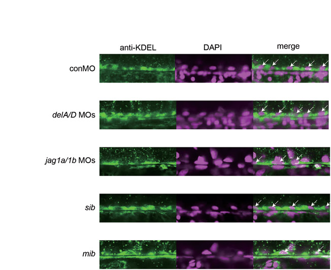
Fig. S7. Mind bomb-Jagged 1 signalling is required for the development of non-vacuolated cells with abundant rough ER. The number of non-vacuolated cells stained by an anti-KDEL antibody was reduced in the jag1a/1b-knockdown and mib mutant embryos, but not in deltaA/D-knockdown embryos. The ER was stained with an anti-KDEL antibody (green), and nuclei were revealed by DAPI staining (magenta). Arrows indicate anti-KDEL-positive non-vacuolated cells. Side views of the ventral regions of the notochord in embryos on day 2 are shown.
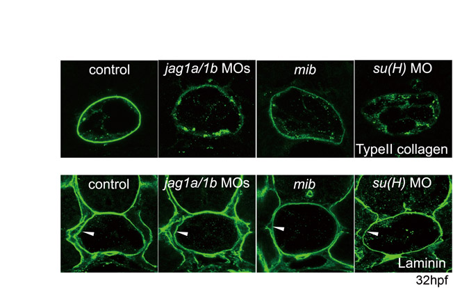
Fig. S8. The medial layer appears to consist of type II collagen. Type II collagen was detected external to the laminin-rich inner layer. The boxed area is enlarged in the lower panels. Green, type II collagen; red, laminin; yellow, merged expression of type II collagen and laminin. Transverse sections through the trunk region of an embryo at 32 hpf are shown.
Fig. S9. Collagen but not laminin protein in the PBM was reduced by the knockdown of Mind bomb-Jagged 1-Notch signalling. Arrowheads indicate laminin expression in the PBM. Transverse sections of the trunk region of embryos at 32 hpf are shown.
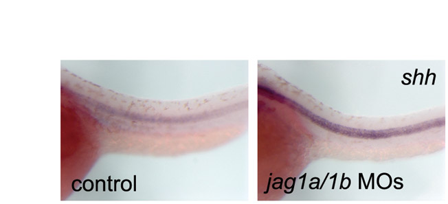
Fig. S10. Expression of shh in Jagged 1-Notch signalling knockdown embryos. The expression of shh did not decrease as the notochord differentiated, in Jag1-Notch signalling knockdown embryos (jag1a/1b MOs). Side views of the trunk region of 32 hpf embryos are shown.
Movie 1. Intercalated notochord cells develop into vacuolated and non-vacuolated epithelial cells. Time-lapse imaging of the notochord cells of an embryo at 21 ss and 32 hpf. Notochord cells were tracked by GFP expression under the promoter of the floating head gene (Flh-GFP). Images were captured every 15 minutes for 14 hours. The movie shows lateral views with anterior to the left.