Files in this Data Supplement:
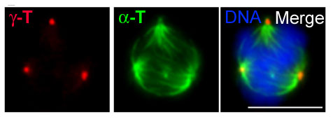
Fig. S1. Expression of stabilized β-cat* induces mitotic defects in a small number of cells. A multipolar spindle with a pair of centrioles at each of three poles is shown in a β-cat*-expressing mitotic MDCK cell with γ-tubulin (red), centrin (green) and DAPI (blue). Scale bar: 10 μm. 1.5±0.01% of mitotic MDCK cells expressing β-cat* have multipolar spindles, whereas only 0.1±0.03% of mitotic parental MDCK cells have multipolar spindles (two experiments, n≥600 each).
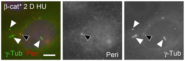
Fig. S2. Extra γ-tubulin puncta are elongated in S-phase-arrested MDCK cells expressing β-cat*. MDCK cells expressing β-cat* were arrested at S-phase for 2 days in hydroyurea (2 D HU) and stained for γ-tubulin and pericentrin. Centrosomes stained for γ-tubulin and pericentrin (black arrowheads), whereas elongated γ-tubulin structures stained for γ-tubulin but not for pericentrin (white arrowheads). Scale bar: 10 m.
Fig. S3. Abnormal elongated centrosomal structures in S-phase-arrested cells expressing β-cat* have no or very little β-catenin or glutamylated tubulin. Magnified centrosomal areas of (A,E) MDCK cells expressing β-cat*, (C) MDCK cell expressing GFP-β-cat*, (B, F) parental HCT116 cells and (D) HCT116 cells expressing GFP-β-cat*. Cells were arrested at S-phase for 3 days with hydroxyurea and immunostained with an antiserum to the C-terminal domain of β-catenin (green: pAb β-cat.-C-term. in A,B) and γ-tubulin (red in A,B) or co-immunostained for GFP (green in C,D), pericentrin (blue in C,D) and γ-tubulin (red in C,D) or co-immunostained for centrin (green in E,F) and glutamylated tubulin (red in E,F). Scale bars: 2 m. Punctate γ-tubulin and β-catenin-positive structures (black arrows in A,B) are probably centrosomes that have been shown before to contain β-catenin, whereas elongated extra γ-tubulin structures have very little β-catenin (white arrows in A,B). GFP-β-cat* localizes to pericentrin-positive centrosomes (dark arrows in C,D) and to only some elongated pericentrin-negative γ-tubulin structures in MDCK (C) and HCT116 (D) cells (white arrows show an elongated structure without GFP-β-cat* in C and with GFP-β-cat* in D, respectively). Centrioles in centrosomes are stained with centrin and glutamylated tubulin (black arrows in F), whereas elongated extra structures (white arrows E,F) are positive for the centriolar marker centrin, but have very little glutamylated tubulin.
Fig. S4. Pre-incubation of anti-Plk4 with antigen reduces Plk4 signal at centrosomes. The ability of the Plk4 antibody to recognize human Plk4 was tested in human embryonic kidney (HEK) 293T cells by co-immunostaining using buffer-preincubated (control) or GST-C-Plk4-preincubated Plk4 antibody. Antigen pretreatment reduces Plk4 immunostaining at centrosomes. Scale bar: 5 m.