Files in this Data Supplement:
Fig. S1. The GFP Protein trap in A2BP1CC00511 reflects the expression and localization of the endogenous protein. (A) A germarium from an A2BP1CC00511 female stained with anti-A2BP1 (red), anti-GFP (green) and 1B1 (blue). The pattern and localization of GFP and A2BP1 are virtually identical. (B) In situ hybridization showing A2BP1 RNA expression.
Fig. S2. Disruption of A2BP1 function leads to extra mitotic divisions resulting in egg chambers with 31 nurse cells. (A,A′) w1118 and (B,B′) A2BP1CC00511 hemizygous egg chambers stained with DAPI and phalloidin. (C) w1118 and (D) A2BP1CC00511/Df(3L)ED4457 ovaries stained with Bam (green) and alpha-Spectrin (red). The Bam expression domain is expanded in the A2BP1CC00511/Df(3L)ED4457 background. (E) Graph showing quantification of the phenotype and suppression by a bam-null mutation. (F) Introduction of an encoreq4 mutation into the A2BP1CC00511/Df(3L)ED4457 background results in the persistence of fusome material in egg chambers. A2BP1CC00511/Df(3L)ED4457 ovaries and (G) encoreq4/+; A2BP1CC00511/Df(3L)ED4457 ovaries stained with 1B1 (red) and DAPI (blue). (H) Graph showing quantification of the phenotype and enhancement by the encoreq4 mutation.
Fig. S3. Disruption of A2BP1 does not result in overlapping expression between Nanos and Bam. Control and A2BP1e03440 hemizygous germaria stained for Nanos (green) and Bam (red). The early expression of both Nanos and Bam do not appear to be disrupted in A2BP1e03440 mutants. The Bam expression domain appears to be greatly expanded in the A2BP1 mutant background.
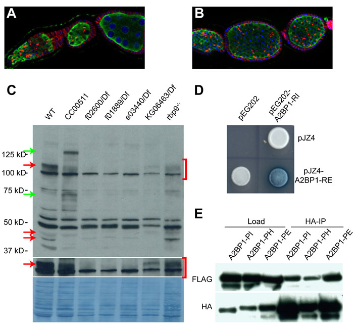
Fig. S4. Different A2BP1 isoforms interact with one another. (A,B) Ovarioles stained for Vasa (green), 1B1 (red) and DAPI (blue). (A) A2BP1KG06463/A2BP1f01889. (B) A2BP1KG06463/ A2BP1e03440. The A2BP1KG06463allele in combination with A2BP1f01889or A2BP1e03440exhibits germ cell counting defects, fusome perdurance past when the organelle is normally degraded and an occasional tumorous phenotype. (C) Western blot showing A2BP1 levels in ovaries from w1118, A2BP1CC00511 homozygotes and various mutant backgrounds, including other A2BP1 alleles and rbp92690. rbp92690 mutants display a tumorous phenotype but express normal levels of A2BP1. The membrane is blotted with polyclonal guinea pig anti-A2BP1 antibody at 1:5000. Red arrows indicate A2BP1 isoforms that exhibit different expression levels in different mutants and green arrows indicate the shifted A2BP1 isoforms due to GFP fusion in the A2BP1CC00511 line. (D) Yeast two-hybrid assay showing the interaction between two different A2BP1 isoforms, RE and RI. (E) Co-immunoprecipitation from S2 cells showing that A2BP1 isoforms bind to themselves.