Files in this Data Supplement:
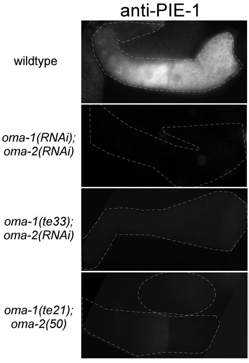
Fig. S1. Immunofluorescence micrographs of anti-PIE-1 staining in the gonad of animals of the indicated genotypes, related to Fig. 3.
Fig. S2. Specificity of GFP::H2Bzif-1 expression pattern, related to Fig. 4. (A) Temporal and spatial expression of GFP::ZIF-1zif-1 is similar to that of GFP::H2Bzif-1. Top panel: Fluorescence micrograph of a wild-type animal expressing GFP::ZIF-1zif-1. Similar to GFP::H2Bzif-1, GFP::ZIF-1zif-1 is repressed in the gonad and early embryos. Middle panel: cul-2(RNAi) results in an increase in GFP::ZIF-1zif-1 level without altering its temporal or spatial location. Bottom panel: Repression in oocytes is released in oma-1(RNAi);oma-2(RNAi) animals. Note cytoplasmic expression, unlike the nuclear expression of GFP::H2B. (B) Repression of GFP::H2Bzif-1 in the pachytene and distal meiotic regions is dependent on GLD-1. DIC images (left) and fluorescence micrographs (right) of animals expressing GFP::H2Bzif-1 in the indicated genetic backgrounds. Ectopic expression of GFP::H2Bzif-1 was observed in gld-1(q485) animals (top and middle rows). In most animals, the ectopic expression was observed only in a tight zone of nuclei adjacent to the spermatheca (top row). It has been shown that a loss-of-function mutation in gld-1 results in meiotic nuclei exiting meiosis and reentering mitosis in the proximal gonad (Francis et al., 1995). The ectopic expression observed in gld-1(q485) is consistent with the expression of GFP::H2Bzif-1 in the mitotic germline stem cells (Fig. 4B). In some gld-1(q485) animals, ectopic expression was observed in a broader zone within the proximal gonad (middle row). In gld-1(q485) animals, OMA proteins are expressed precociously (Lee and Schedl, 2001) (T.G.-O. and R.L., unpublished observations), which could repress translation of Ppie-1-gfp::h2b-UTRzif in the absence of GLD-1. We showed that depletion of oma-1 and oma-2 in gld-1(q485)gld-1(q485);oma-1(RNAi);oma-2(RNAi) animals resulted in the expression of GFP::H2Bzif-1 throughout the gonad in most animals (bottom row). (C) OMA proteins do not repress the translation of a reporter containing the glp-1 3′ UTR in oocytes. DIC (left) and matching fluorescence micrographs (right) of animals expressing GFP::H2Bglp-1 or GFP::H2Bzif-1in wild-type (top row) or oma-1(RNAi);oma-2(RNAi) animals (bottom row). The GLP-1 protein has a similar temporal and spatial expression pattern as GFP::H2Bzif-1 and this is recapitulated with GFP::H2Bglp-1 (Evans et al., 1994; Merritt et al., 2008). Repression of glp-1 in oocytes is not dependent on OMA proteins. Scale bars: 30 µm.
Fig. S3. Deletion analyses of the zif-1 3′ UTR in vivo, related to Fig. 5. Fluorescence micrographs of animals expressing GFP::H2Bzif-1 under the control of zif-1 3′ UTR with specified regions deleted (Fig. 5A). Deleting regions I or IV individually did not result in derepression in the gonad. Deleting regions II or III individually resulted in weak expression of GFP::H2Bzif-1. Scale bars: 30 µm.