Do Liposomal Apoptotic Enhancers Increase Tumor Coagulation and End-Point Survival in Percutaneous Radiofrequency Ablation of Tumors in a Rat Tumor Model?
© RSNA, 2010
Supplemental Figures
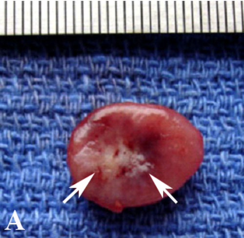 |  |
 | Figure E1: A–C, Gross pathologic specimens of RF ablation–induced coagulation in different treatment groups. Compared with, A, RF ablation alone at 24 hours, B, paclitaxel–RF ablation substantially increased the tumor coagulation (arrows) from 138.8 mm2 ± 12.7 to 488.4 mm2 ± 48.7 (P < .001). C, Tumor coagulation (arrows) was further improved with triple therapy (paclitaxel–RF ablation–doxorubicin), from 488.4 mm2 ± 48.7 to 595.9 mm2 ± 80.9 (P = .016). Scale is in millimeters. |
 |  |  |
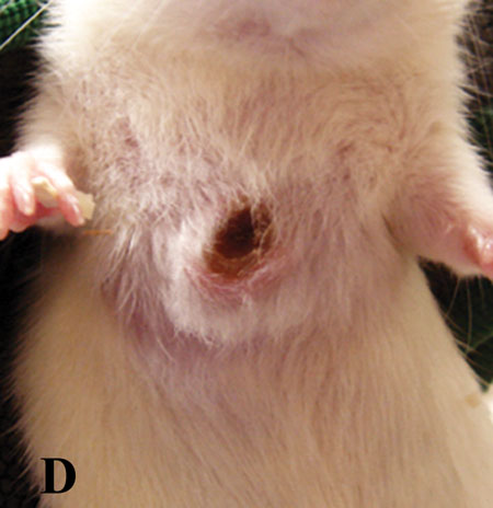 |  |  |
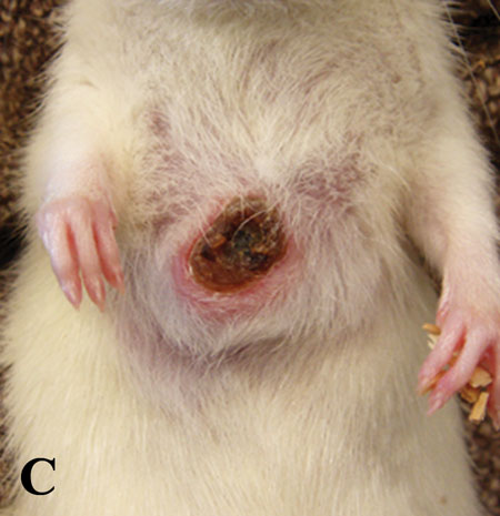
Figure E2: Gross tumor progression and local control in animals treated with combined RF ablation and IV doxorubicin and paclitaxel. A,The initial tumor on day 0 (prior to any treatment) measures 15 × 12 mm. B,At day 20, there were signs of tumor necrosis. C, At day 41, tumor size decreased to 13 × 11 mm with necrosis. D, At day 51, tumor size decreased to 10 × 9 mm with necrosis. E, At day 77, only granulation tissue can be seen. At day 90 (lower right image), no evidence of tumor can be seen, a fact verified at autopsy.
|