Kre6 Protein Essential for Yeast Cell Wall β-1,6-Glucan Synthesis Accumulates at Sites of Polarized Growth
Supplemental Data
Files in this Data Supplement:
Supplementary Figures 1-4 (.pdf, 2.8 MB)
- Supplementary figures.
Figure 6 (.jpg, 313 KB)
- FIGURE 6. Immunofluorescent images of Kre6 in comparison with the SV/PM-marker Snc1.
Figure 2 (.jpg, 73 KB)
- FIGURE 2. Specificity of rabbit anti-Kre6 antiserum.
Figure 4 (.jpg, 229 KB)
- FIGURE 4. Sucrose density gradient fractionation of Kre6-containing compartments.
Figure 1 (.jpg, 284 KB)
- FIGURE 1. Localization of wild-type Kre6 and the tagged Kre6-3HA proteins expressed from the chromosomal genes.
Figure 5 (.jpg, 286 KB)
- FIGURE 5. Immunofluorescent images of Kre6 in Comparison with the ER-marker Lip1.
Figure 8 (.jpg, 185 KB)
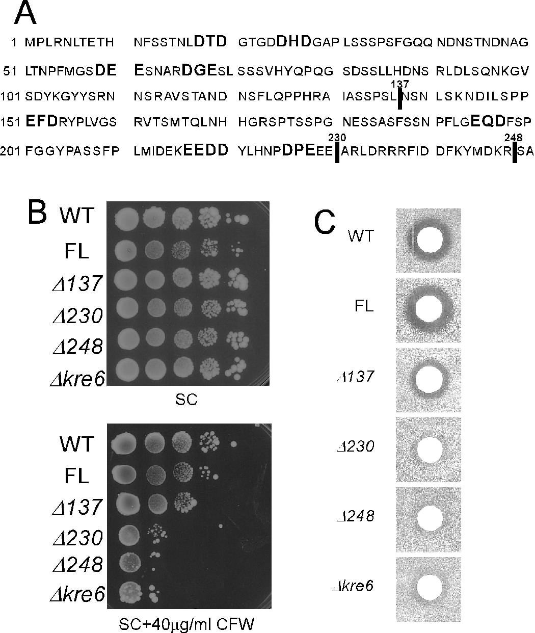
- FIGURE 8. Deletions including possible ER-exit motifs in the cytoplasmic domain and the biological activity of Kre6.
Figure 3 (.jpg, 77 KB)
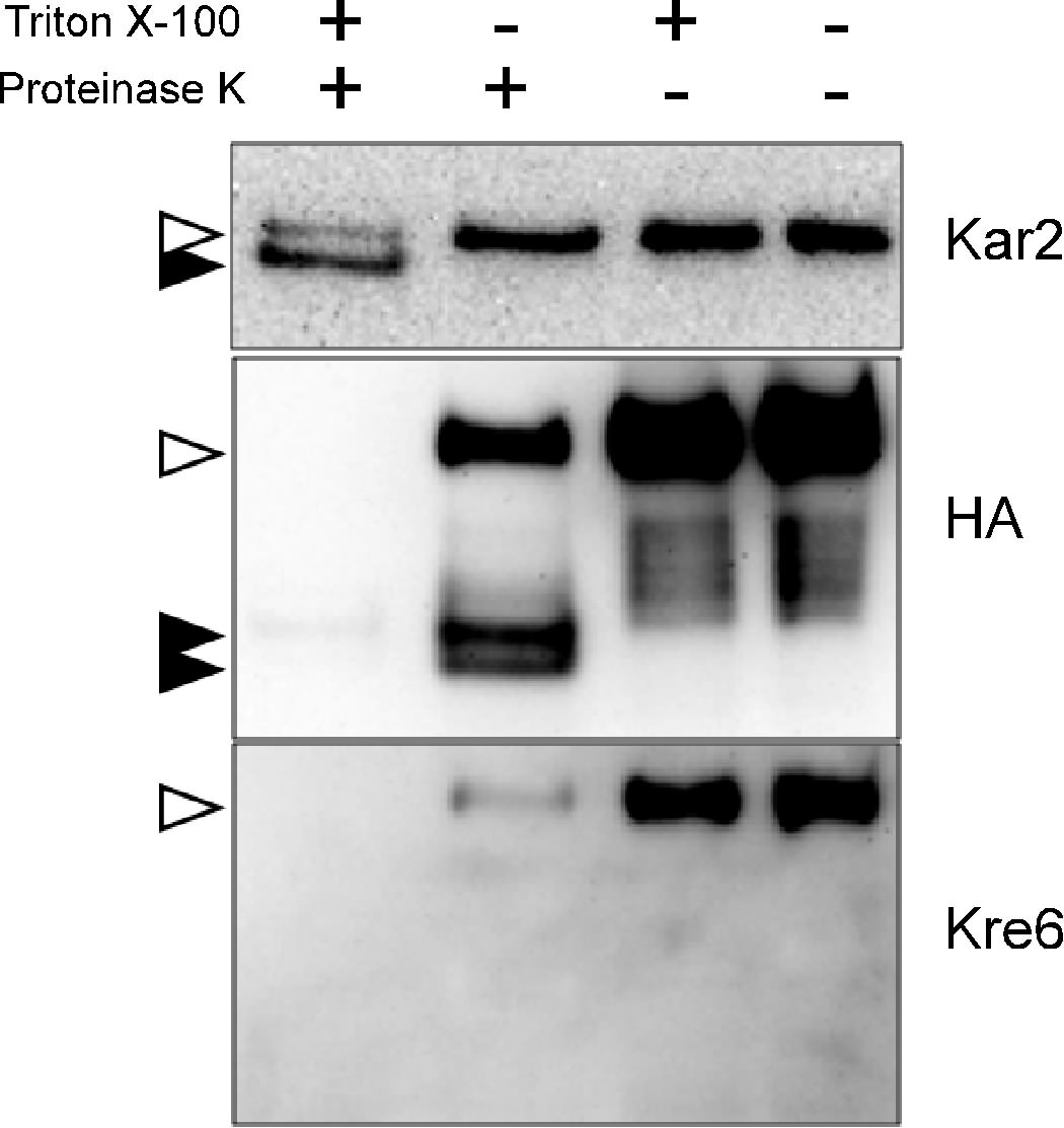
- FIGURE 3. Topology analysis of Kre6-3HA protein by protease protection assay.
Figure 7 (.jpg, 9.0 MB)
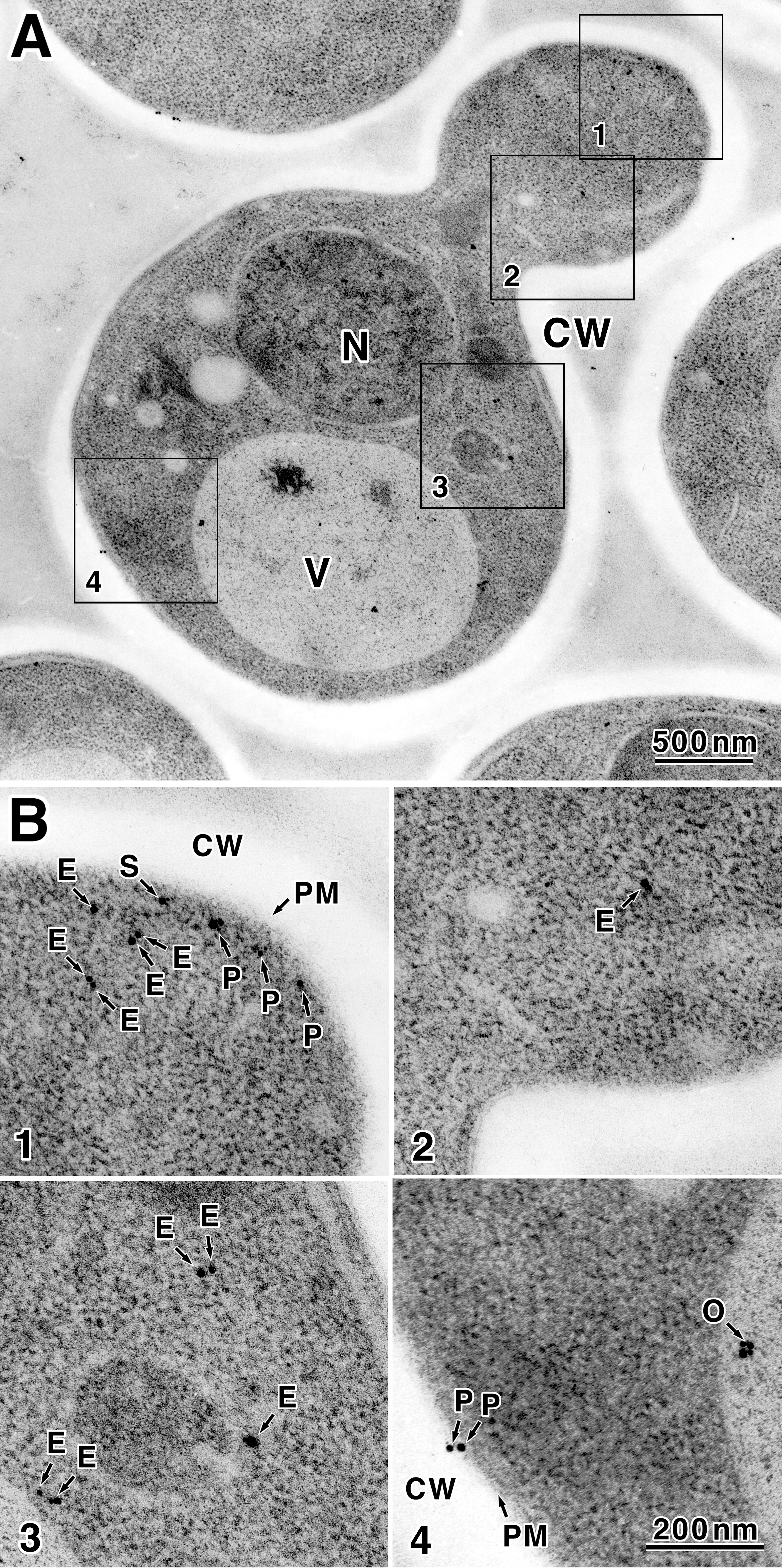
- FIGURE 7. Localization of Kre6-3HA decided by immunoelectron microscopic observation.