Files in this Data Supplement:
Fig. S1. Transient links between the primordial hindbrain channels and basilar artery. (A) Confocal images of the PHBC, BA and transient endothelial connections between them (red arrows) in the hindbrain of Tg(fli1a:EGFP)y1 animals at 24, 33 and 42 hpf. Dorsal view, rostral is to the left. (B) Quantification of the number of PHBC-to-BA connections present between 24 and 45 hpf (n=10 animals). Error bars indicate s.e.m. Scale bar: 50 µm.
Fig. S2. Central arteries project through the center of the rhombomeres. (A-D) Confocal images of the hindbrain from a single Tg(kdrl:mCherry-CAAX)y171; Tg(pax2a:GFP)e1 double-transgenic embryo at 30 hpf (A,C) and 48 hpf (B,D), showing mCherry-positive blood vessels (A,B) or mCherry-positive blood vessels plus GFP-positive rhombomeres 3 and 5 (r3 and r5) and hindbrain neurons (C,D). The first pair of CtAs (white arrows) project dorsally through the middle of r3 (yellow brackets show r3 and r5), eventually linking directly with the BA. More posterior CtAs can be seen projecting through the center of r4, r5 and r6 (white arrowheads). Dorsal-lateral view, rostral to the left. Scale bar: 50 µm.
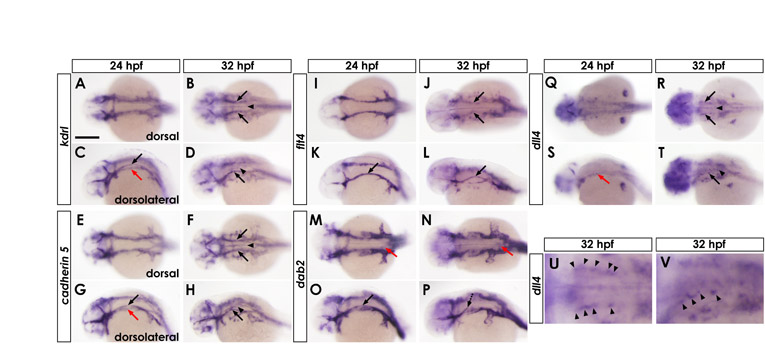
Fig. S3. Expression patterns of endothelial markers. (A-V) Whole-mount in situ hybridization of 24 hpf (A,C,E,G,I,K,M,O,Q,S) and 32 hpf (B,D,F,H,J,L,N,P,R,T,U,V) wild-type zebrafish embryos, probed for kdrl (A-D), cadherin 5 (E-H), flt4 (I-L), dab2 (M-P) or dll4 (Q-V). Dorsal view (A,B,E,F,I,J,M,N,Q,R,U) and dorsolateral view (C,D,G,H,K,L,O,P,S,T,V), rostral is to the left. U and V show magnified portions of the hindbrain from R and T, respectively. Expression of kdrl and cadherin 5 is detected in cranial vessels including PHBC (black arrows), BA (arrowheads), and LDA (red arrows). Expression of flt4 and dab2 is detected in venous vessels including PHBCs (black arrows), although reduced at later stages (dashed black arrows). Weak expression of dab2 is also detectable in LDA (red arrows in M,N). Expression of dll4 is detected in arterial vessels, including BA (arrowheads in R,T) and LDA (red arrow in S). Punctate expression of dll4 is also detectable in central areas of the PHBC (black arrows in R,T, and arrowheads in magnified images in U,V). Scale bar: in A, 200 µm for A-T and 62 µm for U,V.
Fig. S4. VEGF signaling is required for hindbrain vascular patterning. (A,B) Confocal images of the PHBC, BA and CtA in the hindbrain of 30 hpf Tg(kdrl:mCherry-CAAX)y171 animals treated from 24-32 hpf with either control DMSO carrier (A) or the VEGFR inhibitor SU5416 (B). PHBC sprouting defects are present and BA fails to form (arrowheads) in embryos treated with SU5416 after PHBC assembly. PHBC size (yellow brackets) and CtA sprouting (arrows) are also affected in SU5416-treated embryos. Scale bar: 50 µm.
Fig. S5. Expression domains of cxcl12b and cxcr4a in sections. (A,B) Whole-mount in situ hybridization of 32 hpf wild-type zebrafish embryos probed for cxcl12b (A) or cxcr4a (B). Same images as Fig. 4B,H. (C,D) Transverse sections of A (C) and B (D) at the otic vesicle level in the hindbrain (yellow lines in A and B). Expression of cxcl12b is detected in the hindbrain floor plate (black arrows) and the pharyngeal endoderm (black arrowheads). Expression of cxcr4a is detected in the PHBC (blue arrows), BA (red arrows), LDA (red arrowheads) and CtA (purple arrowhead). Lack of PHBC staining on the left side of this section reflects its discontinuous punctate expression pattern (staining on this side is present in other sections). N, notochord; HB, hindbrain; OV, otic vesicle.
Fig. S6. Defects in lateral dorsal aorta formation in cxcl12b and cxcr4a morphants. Lateral dorsal aorta formation is incomplete in cxcl12b and cxcr4a morphants, with gaps between the rostral and caudal portions of these vessels in the head. (A-D) Confocal images of the head vasculature in 24 hpf Tg(fli1a:EGFP)y1 animals injected with control MO (A), 2 ng cxcl12b-MO2 (B), 3 ng cxcr4a-MO2 (C), or 1 ng cxcl12b-MO2 + 1.5 ng cxcr4a-MO2 (D). Dorsal-lateral view. For ease of visualization, the anterior LDA is pseudocolored red, the posterior LDA is pseudocolored green, and veins are pseudocolored blue. Asterisks indicate abnormal gaps in LDA. (E) Quantification of the number of MO-injected embryos with no gaps ('present both') or with gaps in the right ('absent right'), left ('absent left'), or both LDAs ('absent') at 24 hpf. MO dosage is indicated (ng). Scale bar: 50 µm.
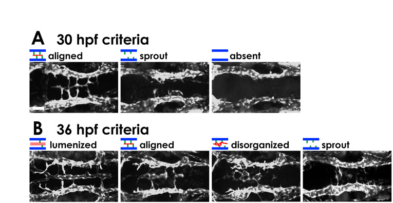
Fig. S7. Phenotypic categories in morphants. (A,B) Typical confocal images of each of the categories of hindbrain vascular phenotype in 30 hpf (A) and 36 hpf (B) MO-injected Tg(fli1a:EGFP)y1 embryos. Dorsal view, rostral to the left. Scale bar: 50 µm.
Fig. S8. Relationship between central artery and basilar artery phenotypes in cxcr4a morphants. Embryos with more severe BA formation defects subsequently reveal more severe defects in CtA growth towards, and connection to, the BA. (A) Transmitted light image (left) shows the region of the hindbrain imaged in the adjacent panels. The remaining panels show typical confocal images of the three categories of r3 CtA phenotype in 42 hpf MO-injected Tg(fli1a:EGFP)y1 embryos. Embryos were first classified at 36 hpf based on their BA phenotype and then reclassified at 42 hpf based on their r3 CtA phenotype. 'Link in r3′, 'midward' and 'dorsalward' mean that the CtA in r3 is already linked with the BA (normal for this stage), fully extended dorsally and turned medially or ventral-medially, and extending dorsalwards but not turning medially, respectively. (B) Quantification of 36 hpf BA phenotypes and subsequent 42 hpf r3 CtA phenotypes in the same animals. The number of embryos analyzed for each MO injection (N) and the 36 hpf BA phenotype class is shown in each box, along with the percentages of these embryos that subsequently fall into each of the three CtA phenotypic classes. Scale bar: 50 µm.
Fig. S9. PI3K signaling is required for HUVEC migration in vitro and for PHBC migration in vivo. (A) SDF-1α-induced dose-dependent migration of HUVECs after a 5 hour incubation, measuring the number of transwell-migrated cells. (B) HUVEC transwell migration in response to 100 ng/ml SDF-1α (column 2) and after pretreatment with either 1 mM AMD3100 (column 4) or 10 mM LY294002 (column 6). Shown (A,B) are the mean (with s.e.m.) obtained by measuring eight randomly selected fields of transwell-migrated cells. (C,D) Confocal images of PHBCs and BA in the hindbrain of 32 hpf Tg(fli1a:EGFP)y1 animals treated from 24-32 hpf with either control DMSO carrier (C) or the PI3K inhibitor LY294002 (D). Dorsal view, rostral to the left. Scale bar: 50 µm.
Movie 1. Two-photon time-lapse imaging of the hindbrain vasculature in a Tg(fli1a:EGFP)y1 zebrafish embryo. The top movie shows BA and CtA formation from 21 to 48 hpf. Endothelial cells sprouting medially from the PHBCs towards the midline form the BA. A separate wave of endothelial cells sprouting dorsally from the PHBCs, beginning at a slightly later stage, form the CtAs. Selected frames from this movie are shown in Fig. 2A and are diagrammed in Fig. 2B. One hundred and sixty-two time points were collected with 10 minutes between time points, ∼27 hours total time. The middle movie is generated from the same time-lapse sequence as the movie at the top, but uses reconstructions of only the dorsal planes of the two-photon image stacks to permit clearer visualization of CtA formation (arrows). One hundred and twenty-one time points from 28 to 48 hpf. The bottom movie is also generated from the same time-lapse sequence as the movie at the top, but uses reconstructions of only the ventral planes of the two-photon image stacks to permit clearer visualization of BA formation (arrowheads). One hundred and twenty-one time points from 28 to 48 hpf. Dorsal view, rostral to the left. Movie rate is six frames/second, or 1 hour elapsed per second of movie time.
Movie 2. Confocal time-lapse imaging of the hindbrain vasculature in a Tg(fli1a:nEGFP)y7 zebrafish embryo. The top movie shows individual endothelial cell nuclei as they contribute to the BA and CtA from 24 to 40 hpf. Endothelial cells sprouting from the PHBCs migrate to and along the midline to form the BA. Selected frames from this movie are shown in Fig. 2C. The bottom movie is generated from the same time-lapse sequence as the movie at the top, but uses reconstructions of only the ventral planes of the two-photon image stacks to permit clearer visualization of BA formation (an example cell is traced by arrows). Dorsal view, rostral to the left. Sixty-five time points were collected with 15 minutes between time points, for ∼16 hours total time. Movie rate is four frames/second, or 1 hour elapsed per second of movie time.
Movie 3. Two-photon time-lapse movie of the hindbrain vasculature in a Tg(fli1a:EGFP)y1 zebrafish embryo. The top movie shows the growth of CtA at the r3 to r6 level from 27 to 40 hpf. Endothelial cells sprout dorsally from the PHBCs to form the CtA. Selected frames from this movie are shown in Fig. 2D. The bottom movie is generated from the same time-lapse sequence as the movie at the top, but uses reconstructions of the proximal planes of the two-photon image stacks to permit clear visualization of only the left PHBC and CtA sprouting on this side at the r3 to r6 level (arrows). Dorsal-lateral view, rostral to the left. Seventy-nine time points were collected with 10 minutes between time points, for ∼13 hours total time. Movie rate is six frames/second, or 1 hour elapsed per second of movie time.
Movie 4. Two-photon time-lapse movie of the hindbrain vasculature in a Tg(fli1a:EGFP)y1 zebrafish embryo. The left movie shows a close-up view of the process of connection between the growing left r3 CtA and the BA, from 30 to 39 hpf. After extending dorsally the r3 CtA sprout turns medially and then ventrally before linking to the BA. Selected frames from this movie are shown in Fig. 2E. The right-hand movie is generated from the same time-lapse sequence as the left movie, but does not use several of the more proximal planes in the reconstructions of the two-photon image stacks so as to eliminate overlapping vessels and permit clearer visualization of the growing r3 sprout (arrows). Frontal-dorsal-lateral view, rostral to the left. Ninety-one time points were collected with 6 minutes between time points, for ∼9 hours total time. Movie rate is ten frames/second, or 1 hour elapsed time per second of movie time.
Movie 5. Confocal image stack of the hindbrain vasculature in control and cxcr4a MO-injected Tg(fli1a:EGFP)y1 zebrafish embryos at 36 hpf. Shown are control (left) and cxcr4a (right) MO-injected Tg(fli1a:EGFP)y1 zebrafish embryos. Dorsal view, rostral to the left. This movie shows each z-section, from ventral to dorsal. The LDA and PHBC/BA are separate in the control embryo, whereas there is an abnormal connection between the LDA and PHBC in the cxcr4a MO-injected embryo (arrows). Seventy-one sections. A reconstructed image generated from these sections is shown in Fig. 6.
Movie 6. Confocal image stack of the hindbrain vasculature in a control and cxcr4a MO-injected Tg(fli1a:EGFP)y1 zebrafish embryos at 36 hpf. Shown are control (left) and cxcr4a (right) MO-injected Tg(fli1a:EGFP)y1 zebrafish embryos. Lateral view, rostral to the left. This movie shows each z-section from medial to lateral. The LDA and PHBC are separate in the control embryo, whereas there is an abnormal connection between the LDA and PHBC in the cxcr4a MO-injected embryo (arrows). Thirty sections. A reconstructed image generated from these sections is shown in Fig. 6.