Files in this Data Supplement:
Fig. S1. Sensitivity of the anti-WRB antibodies. Decreasing amounts of recombinant coiled-coil domain of WRB (WRBcc) were analyzed by SDS-PAGE and immunoblot using anti-WRB antibodies.
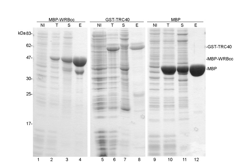
Fig. S2. Purification of recombinant proteins. MBP−WRBcc and MBP were expressed in E. coli and purified by affinity chromatography on amylose resin. GST−TRC40 was expressed in E. coli and purified on glutathione resin. After cell lysis, centrifugation and purification, bacterial lysate before protein induction (NI), total proteins (T), soluble fraction (S) and eluted fraction (E) were separated by SDS-PAGE. Proteins were visualized by Coomassie Blue staining.
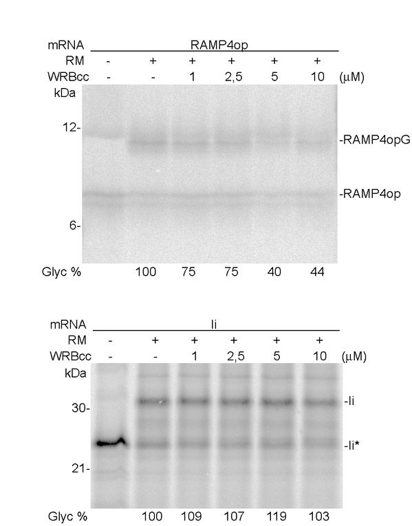
Fig. S3. Effect of WRBcc on the membrane insertion of newly synthesized RAMP4op and invariant chain. RAMP4op and invariant chain (Ii) were translated in vitro in a rabbit reticulocyte lysate in the presence of 35S-labelled methionine and cysteine. Translation was performed in the absence or presence of RMs and increasing amounts of WRBcc as indicated. A quantification of the glycosylated (membrane-inserted) proteins is shown. Proteins were separated by SDS-PAGE and visualized by autoradiography. Ii*: not glycosylated invariant chain.
Movie 1. Colocalization of YFP-TRC40 with WRB-CFP at the ER membrane. WRB-CFP was co-expressed with YFP-TRC40 or with YFP or YFP-TRC40 was expressed alone. Cells were permeabilized with digitonin to release cytosolic content, and the CFP (red), YFP (green) signals or both signals were recorded over time using confocal microscopy.