Files in this Data Supplement:
Fig. S1. Cranial nerve architecture in nhsl1b mutants. Live confocal images of isl1:GFP transgene expression in lateral view in wild-type (A) and nhsl1bfh131 mutant (B) embryos at 48 hours post-fertilization (hpf). Va and Vp, anterior and posterior nuclei, respectively, of the fifth cranial nerve (trigeminal); VII, nucleus of the seventh cranial nerve (facial), which is migrated into r6 in wild-type and is unmigrated in mutant embryos; X, nucleus of the tenth cranial nerve (vagus). Arrows indicate the projection of the facial nerve into the second pharyngeal arch, which is unaffected in nhsl1bfh131 mutants in spite of the failure of the FBM neurons to migrate out of r4. Scale bar: 50 µm.
Fig. S2. Segmental patterning and facial branchiomotor (FBM) neuron differentiation is normal in nhsl1b mutants. (A-D) RNA in situ hybridization with hindbrain markers for rhombomere (r)4 (hoxb1a, blue in A,B); r3 and r5 (krox20; red in C,D); r7 and posterior hindbrain (hoxd4; blue in C,D); and otic vesicles (pax2b, blue in C,D). All of these genes are expressed normally in nhsl1bfh131 mutants (B,D). (E-J) RNA in situ hybridization with motor neuron genes that are required to specify the migratory phenotype: tbx20 (E,F), phox2a (G,H) and tag-1 (I,J). Although FBM neurons fail to migrate caudally in nhsl1bfh131 mutants, they nevertheless express these genes, indicating that their failure to migrate is not due to failure to activate a migratory transcriptional program. Scale bar: 50 µm.
Fig. S3. A splice-blocking nhsl1b morpholino prevents splicing of intron 4. (A,B) A splice-blocking morpholino targeted to the exon 4-intron 4 boundary of nhsl1bfh131 (red bar in A) was injected at the amounts shown. RT-PCR using primers in exon 4 (P1) and exon 6 (P3) amplify two bands corresponding to transcripts with and without exon 5 in wild-type embryos (B). This product decreases in MO-injected embryos. RT-PCR using primers in exon 4 (P1) and intron 4 (P2) amplify a band in MO-injected embryos corresponding to an intron 4-containing transcript, which encodes a truncated protein.
Fig. S4. Expression of nhsl1b homologs nhsa and nhsl1a. (A-D) RNA in situ hybridization of wild-type embryos at the stages shown. nhsa expression overlaps with nhsl1b in somites (bracket in A,C) and at low levels throughout the CNS, but is not upregulated in facial branchiomotor (FBM) neurons. nhsl1a is also expressed at low levels throughout the CNS and is upregulated in rhombomeres (r)5 and r6 (bracket in B,D), in the lens of the eye (asterisk in D) and in cranial ganglia (arrowheads in D), but is not expressed in FBM neurons. hpf, hours post-fertilization. Scale bar: 100 µm.
Fig. S5. Nhsl1b is not required for neuroepithelial apico-basal polarity. (A-D) Confocal images of cross sections at the level of rhombomere (r)4 showing normal apico-basal polarity in the neuroepithelium of 24 hours post-fertilization (hpf) wild-type (A,C) and nhsl1bfh131 mutant (B,D) embryos. ZO-1 marks sub-apical tight junctions of progenitors (A,B) and γ-tubulin marks apical centrosomes (C,D). isl1:GFP marks FBM neurons which accumulate in r4 in nhsl1bfh131 mutants (B,D). Scale bar: 50 µm.
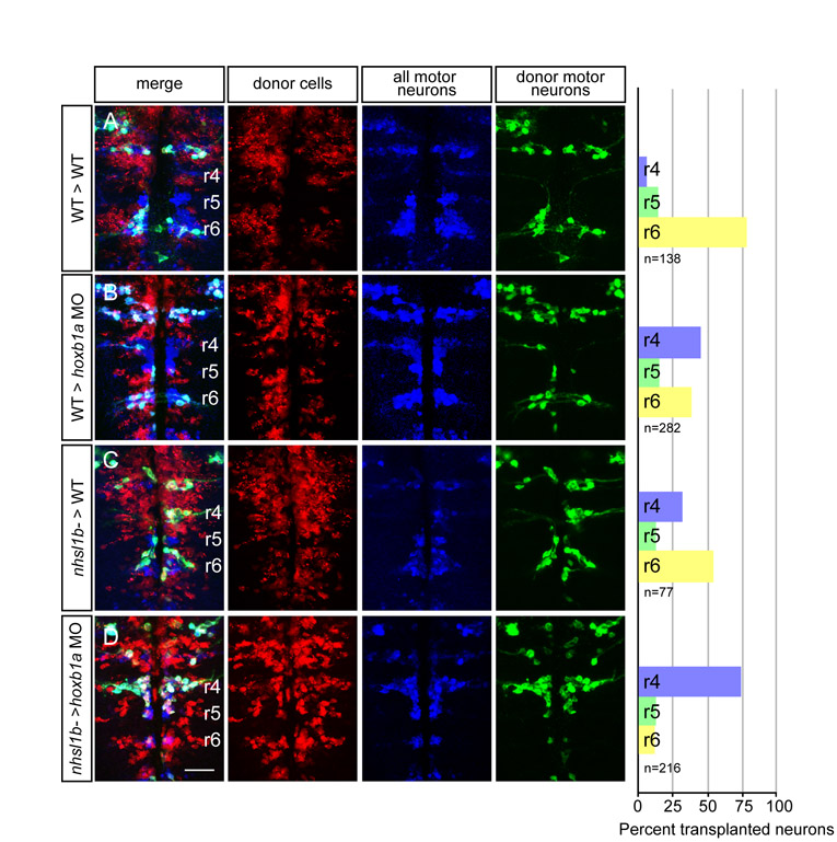
Fig. S6. nhsl1bfh131 mutant facial branchiomotor (FBM) neurons require wild-type FBM neurons for migration. (A-D) Confocal images of fixed mosaic embryos at 48 hours post-fertilization (hpf) with anterior to the top. Lysinated rhodamine dextran marks donor-derived cells (red); anti-Isl1 antibody marks all cranial motor neurons (donor and host-derived, blue); and Tg(isl1:GFP) marks donor-derived motor neurons (green). Histograms on the right indicate the percent of donor-derived FBM neurons in rhombomere (r)4 (unmigrated), r5 and r6 (fully migrated) under the transplantation conditions indicated on the far left, which are written as Donor>Host. n refers to the total number of FBM neurons scored in each condition. hoxb1a morpholinos were used in B and D to prevent host FBM neurons from migrating by a cell-autonomous mechanism. Scale bar: 50 µm.
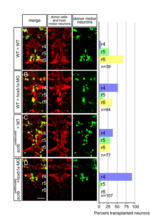
Fig. S7. scribrw468 mutant facial branchiomotor (FBM) neurons require wild-type FBM neurons for migration. (A-D) Live confocal images of mosaic embryos at 48 hours post-fertilization (hpf) with anterior to the top. Lysinated rhodamine dextran marks donor-derived cells (red); Tg(isl1:membRFP) marks host FBM neurons (also red) and Tg(isl1:GFP) marks donor-derived motor neurons (green). Histograms on the right indicate the percent of donor-derived FBM neurons in rhombomere (r)4 (unmigrated), r5 and r6 (fully migrated) under the transplantation conditions indicated on the far left, which are written as Donor>Host. n refers to the total number of FBM neurons scored in each condition. hoxb1a morpholinos were used in B and D to prevent host FBM neurons from migrating by a cell-autonomous mechanism. Scale bar: 50 µm.