Files in this Data Supplement:
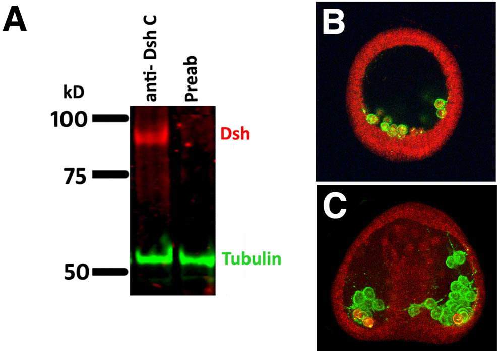
Fig. S1. Western immunoblots. (A) Control western immunoblot for the Dishevelled antibody.An antibody directed against the C terminus peptide of S. purpuratus Dsh recognizes a single band of an egg homogenate; that band is absent if the Dsh Ab is pre-absorbed with a GST fusion Dsh protein. A tubulin antibody is used as a loading control. This western is double-stained with a two-color immunofluorescence kit. (B) NgCAM antibody recognizes PMCs. NgCAM was cloned from L. variegatus and an antibody generated against a GST-fusion protein produced in E. coli. Embryos were injected with rhodamine to provide the red color. (B) Early mesenchyme blastula stage. (C) Late gastrula stage. Embryos are 110 µm in diameter.
Fig. S2. Controls for morpholino knockdowns. (Top) Wnt6 morpholino (MASO) was injected at 300 µm. At 72 hours, controls had skeletons and guts. The Wnt6 MASO-treated embryos had mesenchyme cells but no guts. In the rescue experiments, eggs received 300 µM Wnt6 MASO and mRNA of a Wnt6-GFP construct at 200 ng/µl. The GFP at the 5′ end of the mRNA was added as a control to show the protein was expressed. Five percent were rescued, as shown in the fluorescent embryos on the right, and an additional 20% were partially rescued (n=75). (Bottom) Wnt16 MASO was injected at 150 µM. At 48 hours the MASO-treated embryos had only the beginning of an invaginated gut. Wnt16-GFP was injected into control embryos at 100 ng/µl and had little effect other than increasing the number of pigment cells. Injection of Wnt16-GFP into eggs injected with Wnt16-MASO at 150 µM rescued gastrulation (50% rescued; n=100).
Fig. S3. Controls for Wnt6 overexpression and dnWnt6 in P. lividus. (Top) Overexpression of Wnt6 vegetalizes embryos even when injected at very low concentrations (0.05 µg/µl). (Bottom) dnWnt6 eliminates the gut and endoderm markers at an injected concentration of 0.2 µg/µl. This phenotype is rescued by injection of Wnt6 mRNA at 0.012 µg/µl.
Fig. S4. Maternal S. purpuratus wnt6 in unfertilized eggs. RT-PCR was performed with each of the four primer sets given in the Materials and methods.
Fig. S5. In situ analysis of Wnt6 in the egg. Both in the unfertilized egg (A) and later in the zygote at 1 hpf (B), Wnt 6 mRNA appears ubiquitous. In each case the sense strand controls showed no color at the times development was stopped.