Supplementary material for Wendt et al. (April 3, 2001) Proc. Natl. Acad. Sci. USA, 10.1073/pnas.071051098
Supplemental Movie 1.
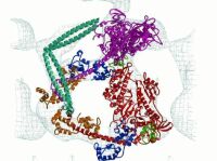
Movie of the smooth muscle heavy meromyosin model fitted to the three-dimensional reconstruction. The initial direction of view is perpendicular to the plane of the crystal (down the z axis of the two-dimensional unit cell). The lipid monolayer would be located below the structure in the initial view. The color scheme for the ribbon diagram is as follows: the heavy chain of the "free" myosin head is magenta and that of the "blocked" head is red. For both myosin heads the converter domain is green, the essential light chains are blue and the regulatory light chains, orange. The movie will rotate about the vertical (y axis) one time with the reconstruction envelope and then rotate once about the x axis without the reconstruction envelope.