Supplementary material for Wendt et al. (April 3, 2001) Proc. Natl. Acad. Sci. USA, 10.1073/pnas.071051098

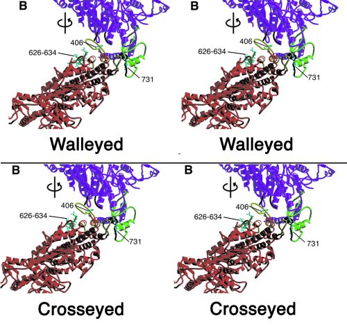
Fig. 7.
Stereoview showing heavy chain–heavy chain interactions. Top pair is for standard stereo viewing; bottom pair is for crosseyed stereo viewing. Same region as Fig. 2A but rotated by ~70° to view within the crystal plane. Color scheme: the heavy chain of the "free" myosin head is magenta and that of the "blocked" head is red. For both myosin heads the converter domain is green, the essential light chains are blue, and the regulatory light chains, orange. In addition, loops involved in heavy chain–heavy chain interactions are in black on the "free" head and in yellow on the "blocked" head. Displayed in cyan in ball and stick format are residues 626–634 of the smooth muscle inhibitory domain and "blocked" head residue 406 and "free" head residue 731.