Files in this Data Supplement:
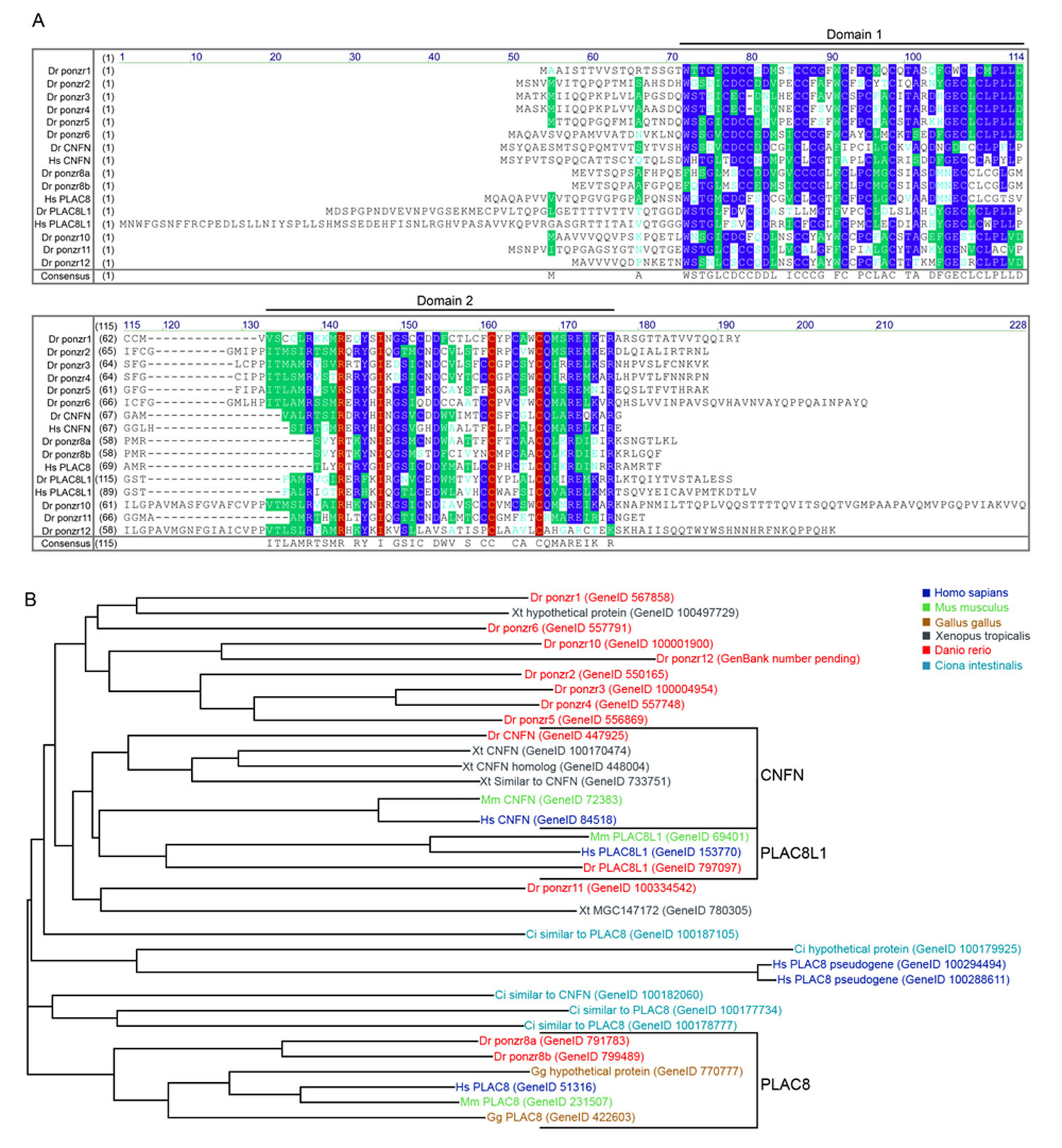
Fig. S1. Sequence alignment of zebrafish and human Ponzr family members. (A) Alignment of the translated amino acid sequence from twelve predicted Ponzr family members found in the zebrafish genome with the three known human Ponzr genes. Domains 1 and 2 are the conserved cysteine-rich domains seen in the Ponzr family and are separated by a variable region. The bottom of the alignment shows the Ponzr family consensus sequence. Blue background with white text shows the consensus amino acid from a block of similar amino acids. Green background with white text shows the amino acid with a greater than 50% occurrence in a position. Red background with yellow text shows complete conservation of the amino acid. Cyan text on white background shows the amino acids that are weakly similar to the consensus. Gene IDs were the chosen identifier and are from NCBI 'GENE' webpage (http://www.ncbi.nlm.nih.gov/sites/gquery). (B) Phylogenetic distance tree shows the evolutionary diversity of this family of proteins. The sequences for this tree were attained by NCBI tBLASTn program using the consensus sequence shown in A and altering the algorithm parameters by increasing the expect threshold to 100 and allowing for low-complexity regions. ponzr1 groups with multiple other zebrafish family members to a single Xenopus gene. When comparing ponzr1 with the Xenopus genome, the first hit was to the Xenopus gene 100497729. When comparing the Xenopus gene with the zebrafish genome, we identified many Ponzr family members (ponzr5, 10, 6, 4 and 1) in that branch of the tree with similar E values. The human pseudogenes map to a specific Ciona gene and, when the pseudogenes were compared to the Ciona genome, the gene 100179925 was the first hit, suggesting that these 'pseudogenes' are actually dying Ponzr family members.
Fig. S2. Early intermediate mesoderm markers are not altered in ponzr1 morphants. wt1a expression in uninjected controls at 4S (A) and 8S (C) does not differ from wt1a expression in ponzr1 morphants at 4S (B) and 8S (D). lhx1 expression in uninjected controls at 8S (E) and 12S (I) is unaltered in ponzr1 morphants at 8S (F) and 12S (J). pax2a expression in uninjected controls at 8S (G) and 12S (K) is similarly unaltered in ponzr1 morphants at 8S (H) and 12S (L).
Fig. S3. Pronephric tubule and duct marker Tg(atp1a1a.4:GFP) shows no changes in pronephric patterning in ponzr1 morphants. (A-P) Wild-type Tg(atp1a1a.4:GFP) embryos at 28 hpf (A, dorsal; B, lateral) and 48 hpf (I, dorsal; J, lateral) show fluorescence in the pronephric tubules and ducts. Mismatch MO-injected embryos show normal fluorescence patterns at 28 hpf (C, dorsal; D, lateral) and 48 hpf (K, dorsal; L, lateral). Embryos were injected at 1 ng (E,F) and 1.5 ng (G,H) of ponzr1 MO1 show no differences in the amount and localization of the fluorescence at 28 hpf (E,G, dorsal; F,H, lateral) and 48 hpf (M,O, dorsal; N,P, lateral). This experiment was conducted twice viewing at least 35 embryos for each group.
Fig. S4. Pronephric tubule and duct marker Tg(enpep:GFP) shows no changes in pronephric patterning in ponzr1 morphants. (A-P) Wild-yype Tg(enpep.4:GFP) embryos at 28 hpf (A, dorsal; B, lateral) and 48 hpf (I, dorsal; J, lateral) show fluorescence in the pronephric tubules and ducts. Mismatch MO-injected embryos show normal fluorescence patterns at 28 hpf (C, dorsal; D, lateral) and 48 hpf (K, dorsal; L, lateral). Embryos were injected at 1 ng (E,F) and 1.5 ng (G,H) of ponzr1 MO1 show no differences in the amount and localization of the fluorescence at 28 hpf (E,G, dorsal; F,H, lateral) and 48 hpf (M,O, dorsal; N,P, lateral). This experiment was conducted twice with at least 15 embryos in each group.
Fig. S5. Knockdown efficacy of ponzr1 MOs. (A) Wild-type embryos show a very low level of baseline GFP autofluorescence. This GFP level was defined as 0% and used to standardize between the three independent experiments. (B) Embryos were injected with ponzr1 5′ UTR-GFP capped mRNA. This GFP level was defined as 100% for each experiment to standardize between the three experiments. (C,D) ponzr1 MO1-injected embryos show a significant loss of fluorescence as compared to both ponzr1 5′ UTR-GFP mRNA and mismatch MO at 1 ng (C) and 2 ng (D) doses. (E,F) ponzr1 MO2-injected embryos also show a significantly decreased fluorescence as compared with ponzr1 5′ UTR-GFP mRNA and mismatch MO at both 2 ng (E) and 3 ng (F) doses. (G,H) Mismatch MO-injected embryos show an attenuated knockdown response, demonstrating only a 50% knockdown at both 2 ng (G) and 3 ng (H). (I) Percentage loss of GFP in MO-injected embryos. ponzr1 MO1 and MO2 are significantly different from both mismatch MO and ponzr1 5′ UTR-GFP (***P<0.001).
Fig. S6. Loss of posterior four pharyngeal arches in ponzr1 morphants. (A) Wild-type larvae at 5 dpf show cartilage staining in 6 pharyngeal arches with the posterior four highlighted in brackets. (B) ponzr1 MO-injected larvae show a loss of the posterior four pharyngeal arches (brackets). (C) Mismatch MO-injected larvae maintain their arches (brackets). (D) This loss of pharyngeal arch cartilage staining is significantly different in the ponzr1 MO1-injected larvae as compared with both wild type and mismatch MO-injected larvae (***P<0.001).
Fig. S7. ponzr1 rescues anterior noitb21−/− mutant phenotype. (A) Mating heterozygous noitb21+/− adults resulted in a subset of embryos with expanded posterior wt1a expression at 24 hpf similar to that described by Majumdar et al. (Majumdar et al., 2000). Injecting 75 pg or 150 pg ponzr1 mRNA into embryos from a heterozygous noitb21+/− incross resulted in a significant reduction of embryos with expanded wt1a expression. **P<0.01. (B) Injecting 75 pg or 150 pg of ponzr1 mRNA into embryos from a heterozygous noitb21+/− incross showed no significant difference in the posterior truncation of cdh17 expression near the cloaca.
Movie 1. Glomerular blood flow in wild-type larvae at 3 dpf. Tg(wt1b:EGFP) was used to focus the microscope and a circle has been placed to help orient where the glomerular capillaries in the Tg(gata1:dsRed) should be. The microscope was switched to an RFP filter and blood flow can be seen within the circle. The bright line of blood cells through the center of the circle is the aorta. The cells moving through the glomerular capillaries can be seen when looking at the outer part of the circle.
Movie 2. Glomerular blood flow in mismatch MO-injected larvae at 3 dpf. This movie shows the Tg(wt1b:EGFP) that was used to focus onto the glomerulus. The fluorescence was then changed to Tg(gata1:dsRed) and the circle shows the area of glomerular flow. The blood moving through the dorsal aorta can be seen running through the middle of the circle. Blood flowing through the glomerular capillaries is seen between the edge of the circle and the aorta.
Movie 3. Loss of glomerular blood flow in ponzr1 MO-injected larvae at 3 dpf. This movie shows the Tg(wt1b:EGFP) that was used to focus onto the glomerulus. The circle shows the area of the glomerulus. The fluorescence was then changed to Tg(gata1:dsRed) to look for blood flow. ponzr1 MO1-injected larvae show blood flow through the dorsal aorta, running in the center of the circle. However, no blood flow can be seen moving through the glomerular capillaries.