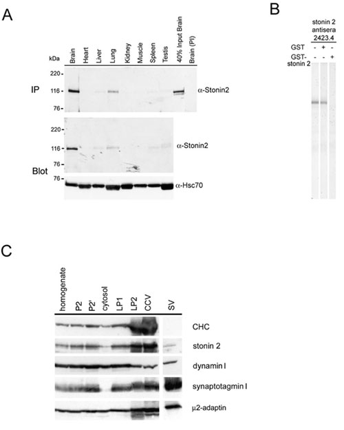
Fig. 6.
(A) Tissue distribution of stonin 2 assessed by multiple-tissue Western blots (Upper) or immunoprecipitation with anti-stonin 2 antisera (Lower). (B) Western blot of rat brain homogenate (50 m g of protein) using stonin 2-antisera preincubated with (+) or without (–) GST or GST-stonin2-NT (5 m g). (C) Subcellular fractionation (30 m g per lane) of pig brain nerve terminals prepared according to Maycox et al. (1). P2, P2', crude and washed synaptosomes; LP1 and LP2, low- and high-speed pellets from lysed synaptosomes; CCV, clathrin-coated vesicles. Purified synaptic vesicles (SV) are shown for comparison. Immunoblots were decorated with antibodies against clathrin heavy chain (CHC), stonin 2, dynamin I, synaptotagmin I, and the m 2 subunit of AP-2.1. Maycox, P. R., Link, E., Reetz, A., Morris, S. A. & Jahn, R. (1992) J. Cell Biol. 118, 1379-1388.