Files in this Data Supplement:
Fig. S1. Gene product from the Tcf3ΔN knockin allele. (A) Tcf/Lef−β-catenin luciferase reporter assay showing effects of expressing combinations of β-catenin, Tcf3, Lef1 and Tcf3ΔN gene products by transient transfection in Cos-7 cells. Relative SuperTOPFlash activity is shown compared to Renilla luciferase control for each transfection. Values represent the mean of biological triplicates ± s.d. (B) Reverse transcriptase PCR analysis of Tcf3 5′ region of Tcf3+/+ and Tcf3+/ΔN heart, lung and liver cDNA samples. The KI mutation introduces an EagI site into the PCR-amplified region. EagI-digested RT-PCR amplicons show EagI-containing cDNA only in Tcf3+/ΔN samples. (C) PCR genotyping of a litter of E8.5 embryos from mating two Tcf3+/ΔN mice (top). Western blot shows expression of WT and Tcf3ΔN proteins in Tcf3+/ΔN ESCs (bottom). Increased mobility of Tcf3ΔN protein reflects correct incorporation of the ΔN mutation. (D,E) H&E-stained tissue sections from submandibular salivary gland (D) and liver (E) of WT E18.5 embryos.
Fig. S2. Proliferation, apoptosis and Tcf/Lef expression during limb morphogenesis. (A,A′) Immunofluorescence staining for phospho-histone H3 (green) and nuclei (red) in E10.0 limb buds from WT (A) and KI (A′) embryos. A representative region near the dorsoventral center is shown. (B,B′) Immunofluorescence staining for cleaved caspase 3 (green) and nuclei (red) in E10.0 limb buds from WT (B) and KI (B′) embryos. A representative region near the dorsoventral center is shown. (C-F′) Immunofluorescence staining for Tcf3 (C,C′), Lef1 (D,D′), Tcf1 (E,E′) and Tcf4 (F,F′) in E10.0 limb buds from WT (C-F) and KI (C′-F′) embryos. Solid lines indicate that Lef1 and Tcf1 decrease in expression in KI (D′,E′) embryos and dashed lines indicate regions where Lef1 and Tcf1 expression is similar to WT (D,E).
Fig. S3. Signaling in the KI eyelid. (A-C′) Immunofluorescent staining of eyelids from WT (A-C) and KI (A′-C′) E14.5 embryos. Antibodies detected phosphorylated-ERK (A,A′), phosphorylated-JNK (B,B′) and phosphorylated-c-Jun (C,C′). (D,D′) FITC-phalloidin (green) staining of filamentous actin is abundant in the periderm tip of the eyelid in WT (D), but not KI (D′), eyelids at E15.5.
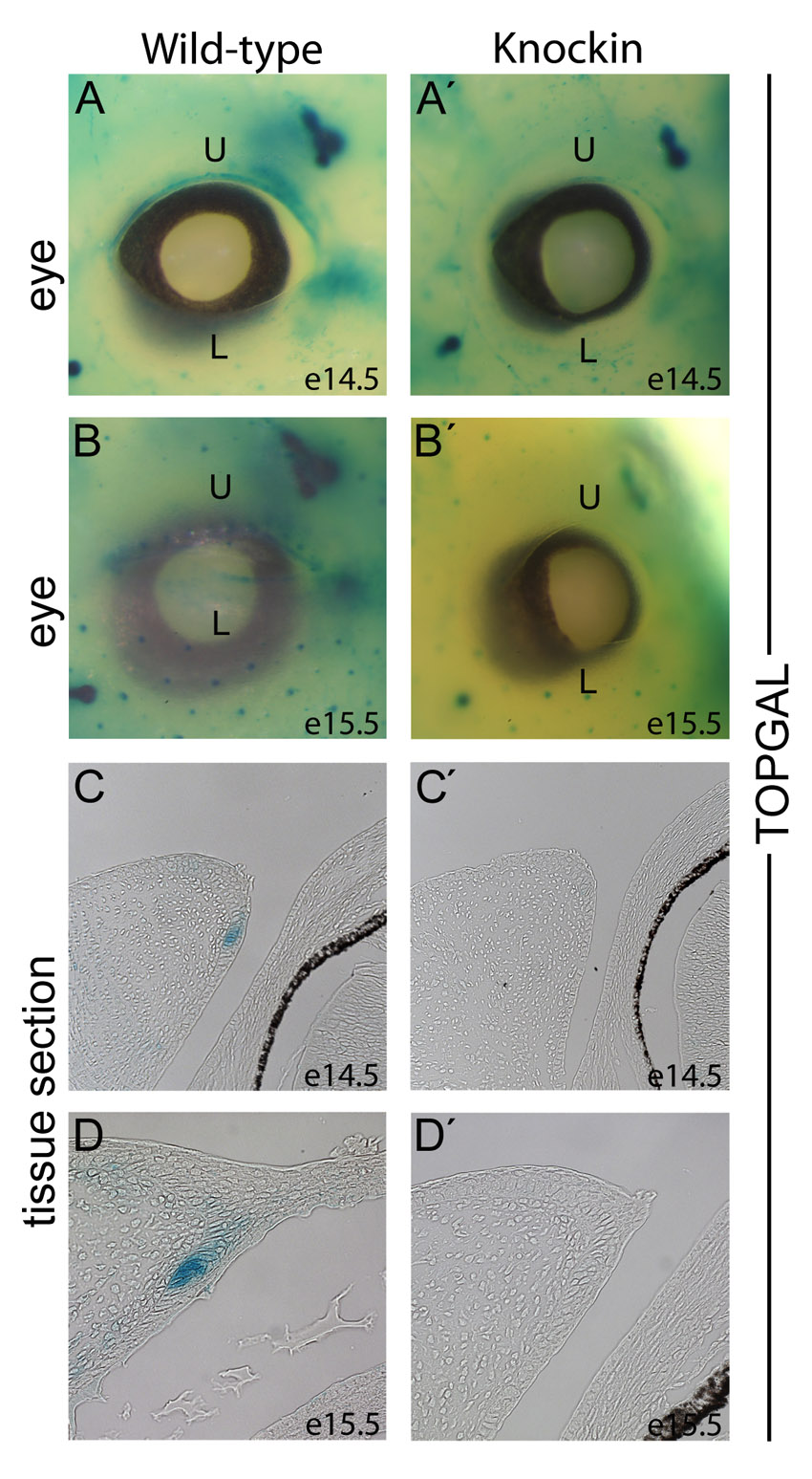
Fig. S4. TOPGal activity in WT and KI eyelids. (A-B′) Whole-mount X-Gal staining of TOPGal-positive WT (A,B) and KI (A′,B′) embryos at E14.5 (A,A′) and E15.5 (B,B′). U, upper eyelid; L, lower eyelid. (C-D′) X-Gal staining of coronal sections through the eye and eyelid of WT (C,D) and KI (C′,D′) embryos at E14.5 (C,C′) and E15.5 (D,D′).
Fig. S5. Tcf/Lef and BAT-Gal expression during eyelid morphogenesis. (A-B′′′) Immunofluorescent staining of coronal section through the eyelid primordia of a BAT-Gal-positive WT embryo at E12.5 (A-A′′′) and E13.5 (B-B′′′). Antibodies specific for Lef1 (red; A,B), Tcf3 (green; A′,B′) and β-galactosidase (blue; A′′,B′′) were used simultaneously on each tissue section. (C,D) Immunofluorescent staining of coronal section through the eyelid primordia of a BAT-Gal-positive WT embryo at E12.5 (C) and E13.5 (D). Antibodies specific for β-galactosidase (red) and Ki67 (green) were used simultaneously on each tissue section. (E,F) Immunofluorescent staining of coronal section through an E14.5 eyelid with an antibody specific for Tcf1 (E) and Tcf4 (F).
Fig. S6. Endogenous Tcf3 represses a Lef1 reporter. (A) −6713/−1 Lef1 reporter plasmid was transiently transfected into WT and KO ESCs. Cells were co-transfected with myc-Tcf3 expression plasmid or its empty vector control and treated with Wnt3a-conditioned media or control conditioned media for 24 hours prior to processing for luciferase activity. Values represent mean of biological triplicates ± s.d. (B) Same experiment as A except the −6713/−879 Lef1 reporter plasmid was used.